Histopathology and Indian Diagnostic Markets
Integrated Diagnostic Workflows
From Blueprint to Details
In pathology, a diagnosis begins with a look at the big picture. A tissue sample stained with hematoxylin and eosin, or , gives pathologists a structural blueprint of the tissue. Hematoxylin stains cell nuclei a purplish-blue, while eosin stains the cytoplasm and connective tissue in various shades of pink. This provides a detailed view of the tissue's architecture, or morphology. Pathologists can see the size and shape of cells, how they are arranged, and whether their structure is normal or disorganized.
However, morphology alone has its limits. Sometimes, cancer cells become so abnormal, or 'poorly differentiated,' that they lose all resemblance to their tissue of origin. A pathologist might see a malignant tumor, but its cells are so distorted that it's impossible to tell if it's a carcinoma (from epithelial cells), a sarcoma (from connective tissue), or a lymphoma (from immune cells). The H&E blueprint shows there's a problem, but it can't identify the culprit.
This is where immunohistochemistry (IHC) comes in. IHC acts as a molecular magnifying glass. Instead of staining structures broadly, it uses antibodies to detect specific proteins, or antigens, on or inside cells. If H&E provides the map of a city, IHC puts the labels on the buildings, telling us which one is a school, a hospital, or a fire station. By identifying a cell's unique protein expression, IHC reveals its identity and function.
Solving Puzzles with Panels
A single IHC stain is rarely enough to solve a diagnostic puzzle. Instead, pathologists employ a 'panel approach,' using a curated set of antibodies to systematically narrow down the possibilities. This is a process of elimination and confirmation. Each marker in the panel acts like a question, and its presence (positive) or absence (negative) provides a critical clue.
Consider a common dilemma: a biopsy from a patient with a known history of lung cancer now shows a tumor in the liver. Is this a new primary liver cancer, or is it a metastasis from the original lung cancer? The treatment for these two scenarios is completely different. H&E staining might show a poorly differentiated adenocarcinoma in the liver, but it can't determine its origin.
By applying a panel, the pathologist can find the answer. Markers like TTF-1 and Napsin-A are typically positive in lung adenocarcinomas, while markers like HepPar-1 and Arginase-1 are specific to liver cells. If the tumor cells stain positive for TTF-1 but negative for HepPar-1, the diagnosis is metastatic lung cancer. This panel approach is indispensable for diagnosing (CUP), where cancer has spread throughout the body but the original site is not obvious. Correctly identifying the primary tumor is critical for selecting the most effective chemotherapy.
Guiding Treatment Decisions
Integrated diagnostics go beyond just naming a cancer. They provide prognostic and predictive information that directly guides treatment. The classic example is in breast cancer. After H&E confirms a diagnosis of invasive ductal carcinoma, an IHC panel is run to check for three key proteins: Estrogen Receptor (ER), Progesterone Receptor (PR), and HER2 (Human Epidermal Growth Factor Receptor 2).
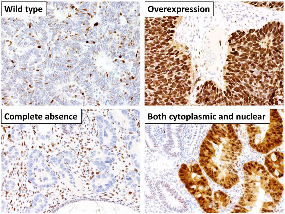
The results of this panel determine the patient's entire treatment course.
-
ER/PR Positive: If the tumor cells express estrogen or progesterone receptors, they are using these hormones to grow. The patient will benefit from hormone therapy, which blocks these receptors or reduces hormone levels.
-
HER2 Positive: If the tumor overexpresses the HER2 protein, it signifies a more aggressive type of cancer. However, this also means the patient is a candidate for targeted therapies like trastuzumab, which specifically attacks HER2-positive cells.
-
Triple-Negative: If the tumor is negative for ER, PR, and HER2, it won't respond to hormonal or HER2-targeted therapies. These patients typically receive chemotherapy.
Molecular subtyping and biomarker testing allow clinicians to tailor therapies to individual tumor characteristics, increasing treatment effectiveness and sparing patients from unnecessary side effects.
This integrated report, combining H&E morphology with IHC-based molecular subtyping, is the foundation of precision oncology. It transforms a general diagnosis into a personalized treatment plan. In Indian oncology and around the world, this synergy doesn't just refine a diagnosis; it directly impacts clinical management, improves patient outcomes, and increases survival rates by ensuring each patient gets the right treatment for their specific cancer.
What is the primary purpose of Hematoxylin and Eosin (H&E) staining in pathology?
A pathologist is examining a tumor in the liver of a patient with a known history of lung cancer. Which of the following IHC panel results would most strongly suggest the liver tumor is a metastasis from the lung?