Heart Valve Leakage Explained
Heart Valve Anatomy
The Heart's One-Way Doors
Your heart is a powerful pump, but its efficiency depends on precise timing and control. A key part of this control system is its four valves. Think of them as one-way doors that open and close with every heartbeat, ensuring blood flows in the correct direction and never backward.
When a heart chamber contracts, it pushes blood forward, forcing a valve open. Once the blood has passed through, that valve snaps shut to prevent any of it from flowing back into the chamber it just left. This coordinated opening and closing happens more than 100,000 times a day.
Heart valves ensure blood moves in a single, forward direction through the heart's four chambers.
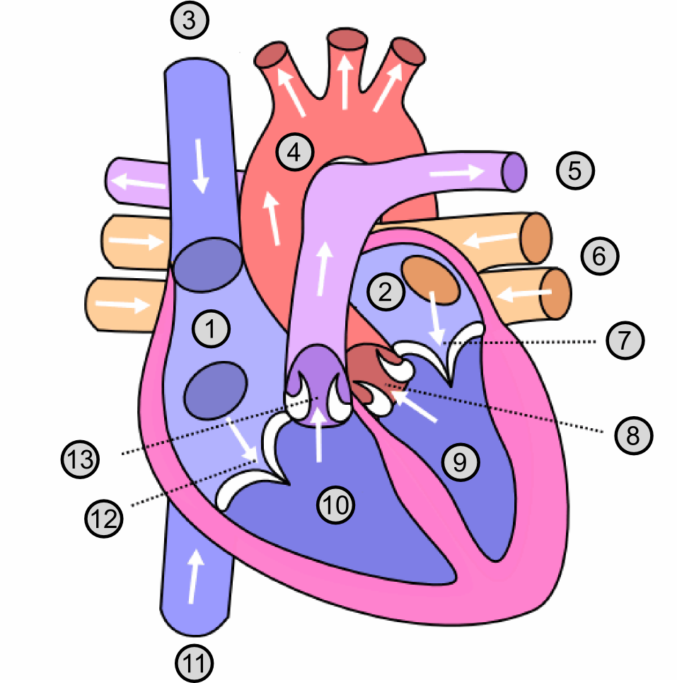
Meet the Four Valves
The heart's four valves can be organized into two types. The first type separates the upper chambers (atria) from the lower chambers (ventricles). The second type controls blood flow out of the heart.
Atrioventricular (AV) Valves These two valves are located between the atria and the ventricles.
- Tricuspid Valve: Positioned between the right atrium and the right ventricle, this valve has three flaps, or cusps. It opens to let oxygen-poor blood from the body pass from the atrium into the ventricle.
- Mitral Valve: Located between the left atrium and the left ventricle, this valve has two cusps. It's also called the bicuspid valve. It opens to allow oxygen-rich blood from the lungs to move from the atrium into the ventricle.
These AV valves have a special anchoring system. They are tethered to the walls of the ventricles by strong, fibrous cords called chordae tendineae, often known as the “heart strings.” These strings attach to small papillary muscles and act like parachute cords, preventing the valve flaps from being forced backward into the atria when the ventricles contract.
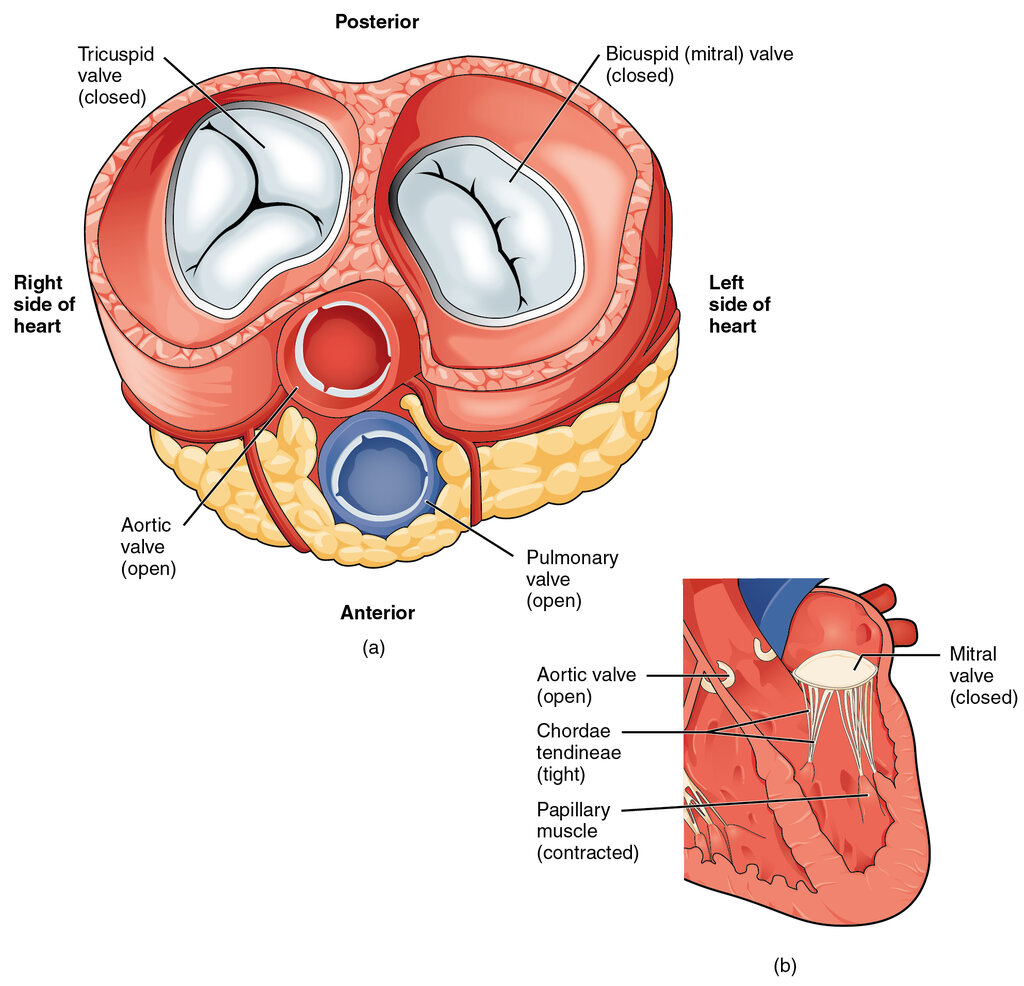
Semilunar (SL) Valves These two valves are located at the exits of the heart, controlling blood flow into the body's major arteries. They have three cusps that look like half-moons, which is where they get their name.
- Pulmonary Valve: This valve sits between the right ventricle and the pulmonary artery. When the right ventricle contracts, it pushes blood through the pulmonary valve and into the pulmonary artery, which carries it to the lungs to pick up oxygen.
- Aortic Valve: This valve is positioned between the left ventricle and the aorta, the body's main artery. When the powerful left ventricle contracts, it forces the aortic valve open, sending oxygen-rich blood into the aorta to be distributed to the rest of the body.
| Valve Name | Location | Number of Cusps |
|---|---|---|
| Tricuspid | Between right atrium and right ventricle | 3 |
| Pulmonary | Between right ventricle and pulmonary artery | 3 |
| Mitral | Between left atrium and left ventricle | 2 |
| Aortic | Between left ventricle and aorta | 3 |
The Path of Blood Flow
The valves work in a perfectly synchronized sequence to guide blood through the heart. It's a two-part cycle that separates oxygen-poor blood from oxygen-rich blood.
-
Right Side (Oxygen-Poor): Blood from the body enters the right atrium. The tricuspid valve opens, letting blood flow into the right ventricle. The ventricle contracts, closing the tricuspid valve and opening the pulmonary valve. Blood is then pumped to the lungs.
-
Left Side (Oxygen-Rich): Blood returns from the lungs to the left atrium. The mitral valve opens, allowing blood to fill the left ventricle. The ventricle contracts, shutting the mitral valve and forcing the aortic valve open. From there, freshly oxygenated blood is sent out through the aorta to the entire body.
While the atrioventricular valves (tricuspid and mitral) are open to let the ventricles fill, the semilunar valves (pulmonary and aortic) are closed to prevent blood from the arteries from flowing back into the heart. Conversely, when the ventricles contract, the AV valves shut while the SL valves open. This perfect coordination creates the familiar “lub-dub” sound of a heartbeat.
Physically trace the path of blood with your finger: from the body (vena cava), to the right atrium, through a valve to the right ventricle, out to the lungs (pulmonary artery), back from the lungs (pulmonary veins), into the left atrium, through a valve to the left ventricle, and finally out to the body (aorta).
Time to check what you've learned about the heart's valves.
What is the primary function of the chordae tendineae, also known as the “heart strings”?
Which valve controls the flow of oxygen-rich blood from the left atrium to the left ventricle?
Understanding the structure and function of each valve is the first step in learning about heart health. Their simple but vital role ensures every part of your body gets the oxygen it needs.