Heart Valve Dynamics
Heart Valve Anatomy
The Heart's Gates
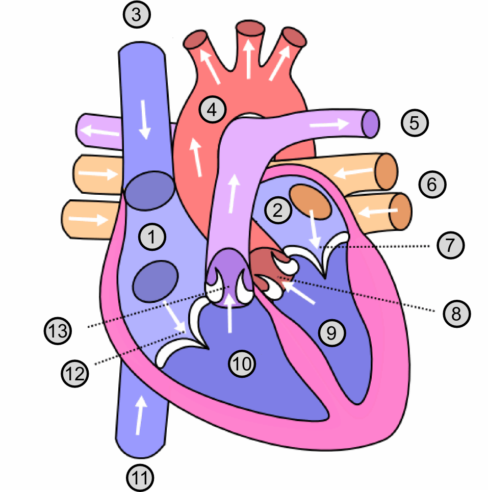
Your heart has four valves that act like one-way doors, ensuring blood flows in a single direction. Without them, blood would slosh backward with every pump, making the circulatory system inefficient. These valves open and close in precise coordination with the heart's contractions, a cycle that repeats about 100,000 times a day.
There are two main types of valves. The atrioventricular (AV) valves sit between the atria (upper chambers) and the ventricles (lower chambers). The semilunar (SL) valves are located at the exits of the ventricles, guarding the pathways into the major arteries.
Atrioventricular Valves
The two AV valves are the tricuspid and mitral valves. They are more complex than the semilunar valves because they have to withstand the high pressure generated when the ventricles contract.
- Tricuspid Valve: Located between the right atrium and right ventricle, this valve has three flaps, or cusps.
- Mitral Valve: Found between the left atrium and left ventricle, this valve has two cusps. It's also known as the bicuspid valve.
leaflet
noun
A flap or cusp of a heart valve. These thin but strong pieces of tissue open to let blood through and seal shut to prevent backflow.
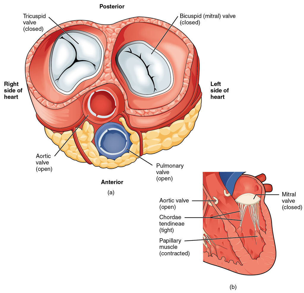
To prevent these valves from being forced backward into the atria during ventricular contraction, they are anchored by a special support system. Thin, fibrous cords called chordae tendineae connect the valve leaflets to small mounds of muscle called papillary muscles, which are part of the ventricle walls. Think of them as the strings on a parachute; they keep the canopy from flipping inside out in the wind.
Semilunar Valves
The semilunar valves are simpler. They don't need chordae tendineae because they don't face the same degree of back pressure as the AV valves. Their name comes from their crescent-moon shape. Each consists of three pocket-like cusps.
- Aortic Valve: This valve sits between the left ventricle and the aorta, the main artery that carries oxygenated blood to the rest of the body.
- Pulmonary Valve: This valve is positioned between the right ventricle and the pulmonary artery, which carries deoxygenated blood to the lungs.
When the ventricles relax, blood in the aorta and pulmonary artery tries to flow back, but it fills the cusps of the semilunar valves, snapping them shut.
The Cardiac Skeleton
All four valves are anchored in a tough, fibrous structure called the cardiac skeleton. This dense ring of connective tissue is crucial for several reasons. It provides a solid foundation for the valve leaflets to attach to, preventing the valve openings from stretching or deforming under pressure.
Beyond structural support, the cardiac skeleton also serves as an anchor point for the cardiac muscle itself. Finally, it acts as an electrical insulator, ensuring that electrical impulses from the atria pass to the ventricles only through the proper conduction pathway, which allows for coordinated contractions.
Let's check your understanding of the heart's valve system.
What is the primary function of the four valves in the human heart?
Which of the following are both considered semilunar (SL) valves?
Understanding this intricate anatomy is the first step in appreciating how the heart works as a perfect, powerful pump.