Heart Anatomy and Cardiology Procedures for Investors
Heart Anatomy
The Heart’s Place in the Body
Your heart is a tireless, muscular pump about the size of your fist. It's nestled safely in your chest, in a space between the lungs called the mediastinum. While we often place a hand over our left chest to feel it, the heart actually sits closer to the center, just slightly tilted to the left. It's protected by your sternum, or breastbone, in the front and your spine in the back.
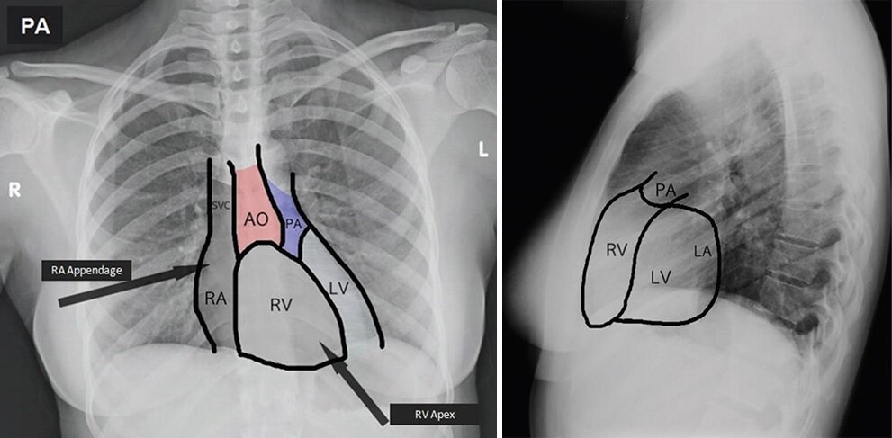
Surrounding the heart is a tough, double-layered sac called the pericardium. Think of it as a protective casing. The pericardium has two main jobs: it anchors the heart in place within your chest, and it prevents the heart from overfilling with blood. A thin layer of fluid between its two layers allows the heart to beat with minimal friction.
The Four Chambers
Inside, the heart is divided into four distinct rooms, or chambers. The two upper chambers are called atria (singular: atrium), and the two lower chambers are the ventricles. A muscular wall called the septum divides the heart into a right side and a left side, creating two separate pumps that work in perfect sync.
The right side of the heart handles deoxygenated blood returning from the body, while the left side manages oxygen-rich blood coming from the lungs.
Here's a breakdown of each chamber's role:
-
Right Atrium: This chamber is the entry point for deoxygenated blood returning from your body. Two large veins, the superior and inferior venae cavae, deliver this blood.
-
Right Ventricle: The right atrium pumps blood down into the right ventricle. From here, the blood is forcefully pumped out of the heart and into the pulmonary artery, which carries it to the lungs to pick up oxygen.
-
Left Atrium: After becoming oxygenated in the lungs, the blood travels back to the heart through the pulmonary veins and enters the left atrium.
-
Left Ventricle: The left atrium moves the fresh, oxygen-rich blood into the left ventricle. This is the heart's strongest and most muscular chamber. It needs that power to pump blood through the aorta, the body's main artery, to supply every organ and tissue with the oxygen it needs.
The wall dividing the two atria is the interatrial septum, and the much thicker wall separating the ventricles is the interventricular septum. These septa are crucial because they prevent oxygen-rich and oxygen-poor blood from mixing.
The Heart's Valves
To keep blood moving in the right direction, the heart has four valves. These valves act like one-way doors, opening to let blood pass through and then snapping shut to prevent it from flowing backward. They are located at the exit of each chamber.
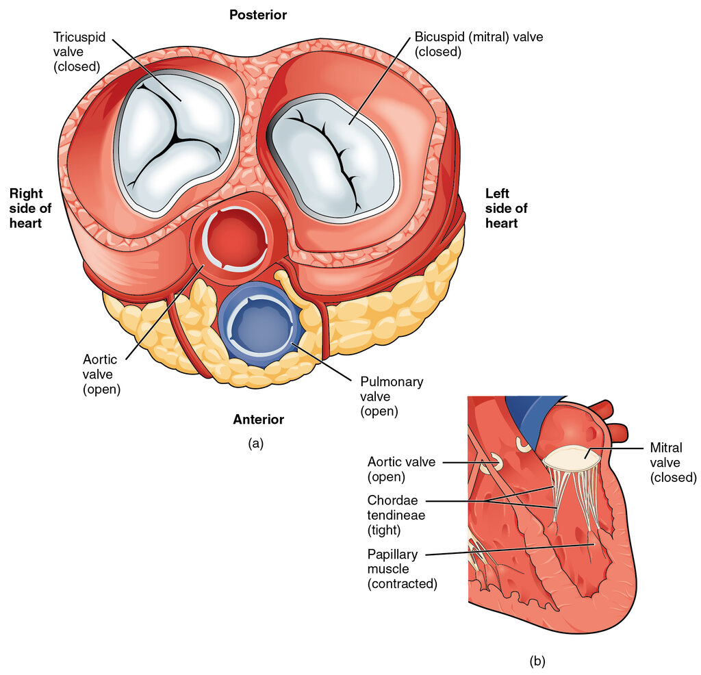
The four main valves are:
- Tricuspid Valve: Located between the right atrium and the right ventricle.
- Pulmonary Valve: Situated between the right ventricle and the pulmonary artery.
- Mitral Valve: Found between the left atrium and the left ventricle.
- Aortic Valve: Positioned between the left ventricle and the aorta.
The coordinated opening and closing of these valves is what produces the familiar "lub-dub" sound of a heartbeat.
Which chamber of the heart is the most muscular and powerful, responsible for pumping oxygenated blood to the entire body?
What is the primary function of the pericardium?