Gonioscopy Essentials
Introduction to Gonioscopy
Looking Around Corners in the Eye
The eye is filled with a clear fluid called aqueous humor, which is constantly being produced and drained. This process maintains a healthy pressure inside the eye. Think of it like a sink with the faucet always on a slow drip; the drain must work properly to prevent an overflow. In the eye, this drain is located in a specific spot called the anterior chamber angle.
This angle is where the cornea (the clear front part of the eye) meets the iris (the colored part). It's a crucial piece of anatomy, but it's impossible to see directly. Due to the way light bends as it passes from the cornea into the air, the angle is hidden from view. It’s like trying to see around a corner without a mirror.
Gonioscopy
noun
A diagnostic procedure that uses a special contact lens to view the eye's anterior chamber angle, the drainage area where aqueous humor exits the eye.
Gonioscopy is the clever solution to this problem. It's a technique that uses a special mirrored or prismatic lens, placed directly on the patient's eye, to act as that mirror. It allows an eye doctor to look around the corner and get a clear view of the drainage system. Its main purpose is to help diagnose and manage glaucoma, a condition often linked to problems with this drainage system.
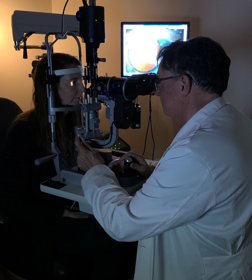
The Drainage Angle Anatomy
To understand why gonioscopy is so important, we need to know what the doctor is looking for. The anterior chamber angle isn't just a simple corner; it's a complex structure made of several key parts working together. The most important of these is the trabecular meshwork, a spongy tissue that acts as the primary filter for the aqueous humor before it exits the eye.
Here's a quick tour of the neighborhood:
- Trabecular Meshwork: The main filter. This is the star of the show in gonioscopy.
- Schlemm's Canal: A circular channel that collects the filtered fluid from the trabecular meshwork and directs it into the bloodstream.
- Scleral Spur: A ring of tissue that serves as an anchor for the iris and ciliary body muscles.
- Ciliary Body: The structure that produces the aqueous humor and also contains the muscle that helps the eye focus.
By examining these structures, an ophthalmologist can determine if the drainage angle is open, narrow, or closed.
The Glaucoma Connection
Glaucoma is a disease that damages the optic nerve, often due to high pressure inside the eye (intraocular pressure). High pressure builds up when the aqueous humor cannot drain properly. Gonioscopy is essential because it helps classify the type of glaucoma.
| Angle Status | Implication for Glaucoma |
|---|---|
| Open Angle | The drainage angle is visibly open, but the trabecular meshwork isn't filtering fluid correctly. This is the most common form of glaucoma. |
| Closed Angle | The iris is physically blocking the trabecular meshwork, preventing fluid from reaching the drain at all. This can be an emergency. |
Knowing whether the angle is open or closed is not just an academic detail; it completely changes the treatment plan. An open angle might be treated with eye drops to improve drainage through the meshwork, while a closed angle might require a laser procedure to create a new path for the iris to move away from the drain.
Without gonioscopy, a doctor would be guessing at the underlying cause of the high pressure. It provides the direct, visual information needed to make an accurate diagnosis and choose the most effective and safest treatment.
What is the primary purpose of gonioscopy?
In the analogy of the eye's fluid system being like a sink, what part does gonioscopy directly inspect?
Gonioscopy provides a direct view into one of the most important functional areas of the eye, turning a potential diagnostic puzzle into a clear clinical picture.