Frontiers in Osteoarthritis PhD Research Design
Adipose Metabolic Hubs
Beyond a Shock Absorber
For a long time, the infrapatellar fat pad (IFP), also known as Hoffa's fat pad, was seen as little more than a cushion for the knee. It sits just below the kneecap, and its primary role was thought to be mechanical: absorbing shock and distributing forces across the joint. While it does serve that function, we now understand the IFP is a complex, metabolically active organ with a powerful influence on the health of the entire knee.
Think of it less like a passive cushion and more like a local command center. The IFP acts as an endocrine organ, producing and secreting a variety of signaling molecules called adipokines. These molecules don't just stay within the fat pad; they are released directly into the synovial fluid, allowing them to communicate with the surrounding cartilage and synovium. This chemical conversation can either help maintain a healthy joint or drive it toward a state of chronic inflammation and degradation, which is a hallmark of osteoarthritis.
The Fat Pad's Chemical Messengers
The adipokines released by the IFP are a mixed bag. Some are protective, while others are destructive, and the balance between them is crucial for cartilage homeostasis. Three of the most studied adipokines in the context of osteoarthritis are leptin, adiponectin, and resistin.
| Adipokine | Primary Role in the Knee Joint |
|---|---|
| Leptin | Mostly pro-inflammatory. Promotes cartilage breakdown by stimulating matrix-degrading enzymes. |
| Adiponectin | Generally anti-inflammatory. Can protect cartilage cells but has complex, dual roles. |
| Resistin | Pro-inflammatory. Associated with increased inflammation and cartilage degradation. |
When the knee joint is healthy, the levels of these adipokines are balanced. However, in an osteoarthritic joint, the IFP often becomes inflamed and dysfunctional. It starts producing higher levels of pro-inflammatory messengers like leptin and resistin, tipping the scales toward cartilage destruction. This constant inflammatory signaling from the fat pad creates a toxic environment for the entire joint.
A Neighborhood of Active Cells
The IFP isn't just a collection of fat cells (adipocytes). It's a diverse ecosystem of cell types, each with a distinct role. Key players include:
- Adipocytes: The primary fat-storing cells, and the main producers of adipokines.
- Macrophages: Immune cells that come in different flavors. M1 macrophages are pro-inflammatory, while M2 macrophages are more involved in tissue repair and resolving inflammation.
- Mesenchymal Stromal Cells (MSCs): These are versatile cells that have the potential to differentiate into various cell types, including cartilage and bone. They can aid in repair but can also contribute to disease.
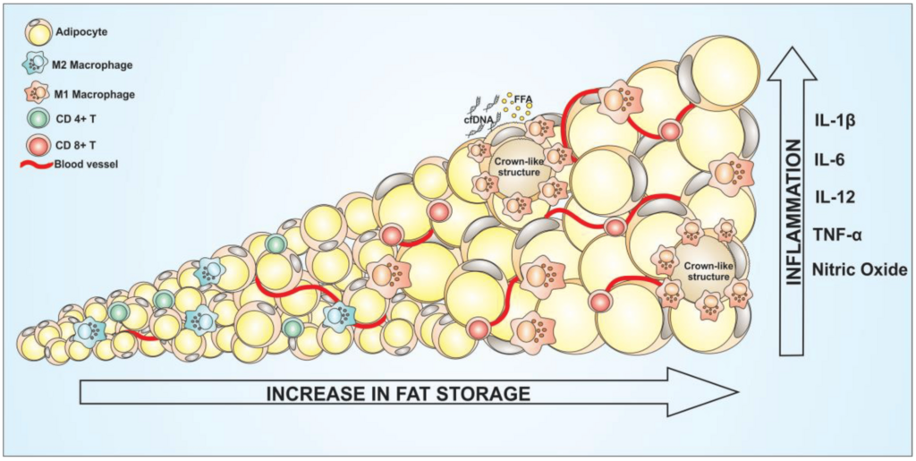
As these cells age or experience stress, they can enter a state known as senescence. Instead of dying, senescent cells stop dividing but remain metabolically active, secreting a cocktail of inflammatory proteins, growth factors, and enzymes. This cocktail is called the senescence-associated secretory phenotype, or SASP.
The SASP from senescent cells in the IFP acts like a continuous alarm signal, perpetuating joint-wide inflammation and preventing any meaningful repair.
This leads to a destructive cross-talk between the joint's tissues. The IFP releases SASP factors and pro-inflammatory adipokines into the synovial fluid. These signals inflame the synovium (the membrane lining the joint), which in turn releases its own inflammatory molecules that attack the cartilage. The damaged cartilage then sends distress signals back, further activating the cells in the IFP and synovium. This vicious cycle is a key driver of osteoarthritis progression, moving it from a disease of simple 'wear and tear' to one of whole-joint inflammation.
The Downward Spiral of Fibrosis
Chronic inflammation and persistent SASP signaling eventually lead to another problem: fibrosis. Fibrosis is the excessive formation of connective tissue, essentially scarring. Within the IFP, this process is driven by activated MSCs and other cells that begin churning out large amounts of collagen and other matrix proteins.
A fibrotic fat pad becomes stiff and dysfunctional. It loses its ability to function as a shock absorber and becomes even worse at regulating its metabolic signaling. The stiffened tissue can also cause mechanical problems, leading to pain from impingement. This scarring isn't just a byproduct of the disease; it's an active part of it, further disrupting the joint's delicate homeostasis and locking it in a diseased state.
Understanding the IFP as a central player in joint health opens new doors for treating osteoarthritis. Instead of just focusing on the cartilage, researchers are now targeting the fat pad, looking for ways to reduce its inflammation, block harmful adipokine signaling, and prevent fibrosis. It’s a shift from a cartilage-centric view to a whole-joint perspective.
Let's review what you've learned about the infrapatellar fat pad's role in the knee.
How has the scientific understanding of the infrapatellar fat pad (IFP) evolved over time?
In an osteoarthritic knee, an inflamed IFP tends to release higher levels of pro-inflammatory messengers like leptin and resistin, tipping the balance toward cartilage destruction.
By seeing the whole picture, we can develop more effective strategies to manage and potentially reverse the effects of osteoarthritis.