Female Reproductive Anatomy and Health
Vulvar Topography
The Protective Outer Layers
The vulvar topography begins with the mons pubis, a pad of adipose tissue situated directly over the pubic bone. This fatty cushion serves as a protective buffer for the bone during sexual activity and everyday movement. Its composition provides both shock absorption and a foundation for the external genital structures.
Extending down from the mons pubis are the labia majora, or outer lips. These two prominent folds of skin are homologous to the scrotum in males and are primarily composed of adipose tissue and a thin layer of smooth muscle. The outer surface is typically pigmented and contains hair follicles, along with both sweat glands and sebaceous glands that produce oils. The inner surface is smooth, hairless, and transitions to a mucous membrane-like texture, reflecting its role as a border to the more delicate internal structures.
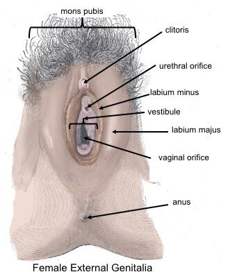
Structurally, the labia majora exhibit significant natural variation in size, prominence, and symmetry. These differences are normal and change throughout a person's life due to factors like age, hormonal shifts, and childbirth. Their primary function is protective, forming a physical barrier that shields the inner vulvar structures, including the urethral and vaginal openings, from pathogens and friction.
Inner Structures and Glands
Tucked within the labia majora are the labia minora, or inner lips. Unlike their outer counterparts, these folds are devoid of adipose tissue and hair follicles. Instead, their core is made of spongy connective tissue containing elastic fibers and a rich network of blood vessels, which contributes to their sensitivity and ability to swell with arousal. They are also rich in sebaceous glands, which provide lubrication.
The labia minora display an even wider array of variation in size, color, and shape than the labia majora. It's common for them to be asymmetrical or to protrude beyond the labia majora. These variations are a normal part of human diversity. The labia minora meet at the top to form the prepuce, or hood, which covers the clitoral glans.
The area enclosed by the labia minora is the —a functionally critical region. This smooth-surfaced space contains the external urethral orifice for urination and the vaginal orifice (or introitus). The vestibule also houses the openings for two important sets of glands: the Skene's glands, located near the urethra, and the Bartholin's glands, situated on either side of the vaginal opening. Both contribute to lubrication, particularly during sexual arousal.
Nerve Supply and Sensitivity
The entire vulvar region is densely innervated, making it highly sensitive to touch, pressure, and temperature. The primary nerve responsible for this sensitivity is the pudendal nerve, a major nerve of the pelvis. It branches extensively to supply the labia, clitoris, and perineum.
This intricate nerve network is composed of different types of nerve endings. Some are specialized for detecting light touch, while others respond to deeper pressure or vibration, contributing to the complex sensations experienced in this area.
The density of these nerve endings is not uniform across the vulva. The labia minora and clitoris, in particular, have a much higher concentration of sensory receptors compared to the mons pubis or the outer surfaces of the labia majora. This anatomical arrangement underscores their central role in sexual response and tactile pleasure. Understanding this complex innervation is crucial for clinical procedures and for appreciating the physiological basis of sexual health.
Let's review the key structures we've covered.
Now, test your understanding of the vulva's anatomy.
What is the primary function of the mons pubis?
Which statement accurately describes a key difference between the labia majora and the labia minora?
By understanding the specific tissues, glands, and nerve networks of the vulva, we gain a deeper appreciation for its dual roles in protection and sensation.