Endoscopic Ultrasound for Biliary and Pancreatic Diseases
Introduction to Endoscopic Ultrasound
A Closer Look Inside
Imagine you want to see what's happening in the room next door. You could press your ear against the wall, but the sound would be muffled. Or, you could drill a tiny hole and put a microphone right inside. Endoscopic ultrasound, or EUS, is like that microphone. It’s a specialized procedure that gives doctors an incredibly detailed, close-up view of your digestive tract and nearby organs like the pancreas and liver.
EUS combines two powerful technologies: an endoscope and an ultrasound. An endoscope is a thin, flexible tube with a camera and light on the end. It allows doctors to look directly inside your body. An ultrasound uses high-frequency sound waves to create images of internal structures. EUS puts a miniature ultrasound probe on the tip of the endoscope. This hybrid tool can be guided down the esophagus into the stomach and the first part of the small intestine, placing the ultrasound just millimeters away from the organs it needs to examine.
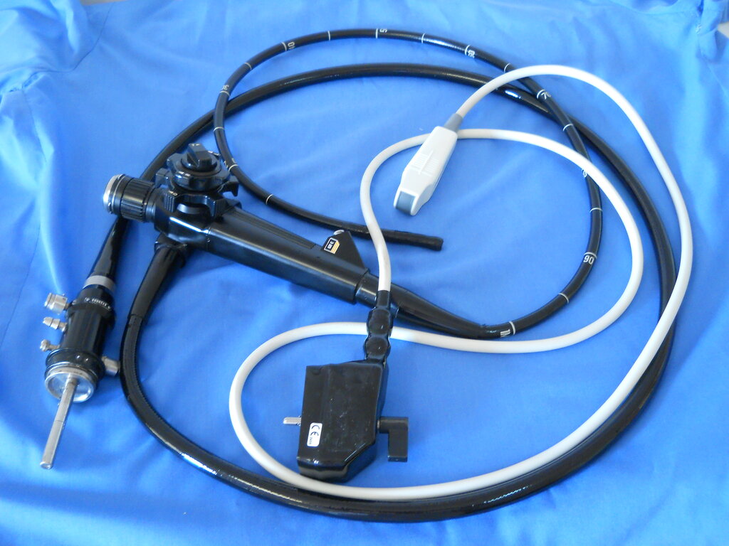
The EUS Procedure
The procedure itself is straightforward from the patient's perspective. Beforehand, you'll be asked to fast for several hours to ensure your stomach is empty, providing a clear view. When you arrive, you'll typically be given a sedative to help you relax and feel comfortable. Most patients sleep through the entire process.
The doctor then gently guides the EUS scope through your mouth, down your esophagus, and into your stomach or small intestine. Once in position, the ultrasound probe sends out sound waves that travel through the wall of your digestive tract and into the surrounding organs. These waves bounce back, creating highly detailed images on a monitor. The whole procedure usually takes between 30 and 60 minutes.
The Advantage of Proximity
So, why use EUS instead of a standard ultrasound, CT scan, or MRI? The answer is proximity. A regular abdominal ultrasound has to send sound waves through skin, fat, and muscle to reach organs like the pancreas. Each layer can distort the signal, resulting in a less detailed image. It's like trying to take a picture of a bird through a foggy window.
EUS bypasses these obstacles. By placing the probe directly against the wall of the stomach or small intestine, it gets an unobstructed, high-resolution view of the pancreas, bile ducts, and lymph nodes. This clarity allows doctors to spot subtle abnormalities, small tumors, or stones that other imaging tests might miss.
The main advantage of EUS is its ability to produce highly detailed images of organs near the gastrointestinal tract by minimizing the distance the sound waves must travel.
This level of detail is crucial for diagnosing conditions and for 'staging' cancers, which means determining their size and whether they have spread to nearby tissues or lymph nodes. Accurate staging is essential for planning the most effective treatment. While EUS is a powerful tool, it's just one of many that doctors use to get a complete picture of a patient's health.