Dynamics of the Human Heart
Cardiac Micro-Anatomy
The Heart's Cellular Machinery
You already know the heart has four chambers that pump blood. But how does this muscle pull off such a perfectly synchronized beat, over and over, for a lifetime? The secret lies in its specialized cells and the unique way they are wired together.
The heart wall is primarily made of cardiac muscle cells, or cardiomyocytes. Unlike the long, straight fibers of your skeletal muscles, cardiomyocytes are branched, shorter, and typically have just one or two nuclei. They are packed with mitochondria, the powerhouses of the cell. This makes sense; the heart is a non-stop engine that requires a colossal and continuous supply of energy.
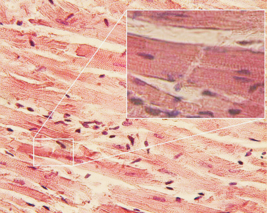
The most crucial feature of these cells isn't visible without high magnification. At the end of each branch, cardiomyocytes connect to their neighbors at specialized junctions called . These are not simple connections; they are complex structures that serve two vital functions: holding the cells together and allowing them to communicate instantly.
One Coordinated Squeeze
Within the intercalated discs, two types of junctions are critical. Desmosomes act like strong molecular rivets, anchoring the cells together. They prevent the cardiac muscle from pulling apart during forceful contractions. Think of them as the physical connection.
Even more important for timing are the gap junctions. These are tiny channels that pass directly from the cytoplasm of one cell to the next. They allow electrical impulses, in the form of ion flow, to travel almost instantaneously across the entire network of cells. This electrical connection is what makes the heart's unique function possible.
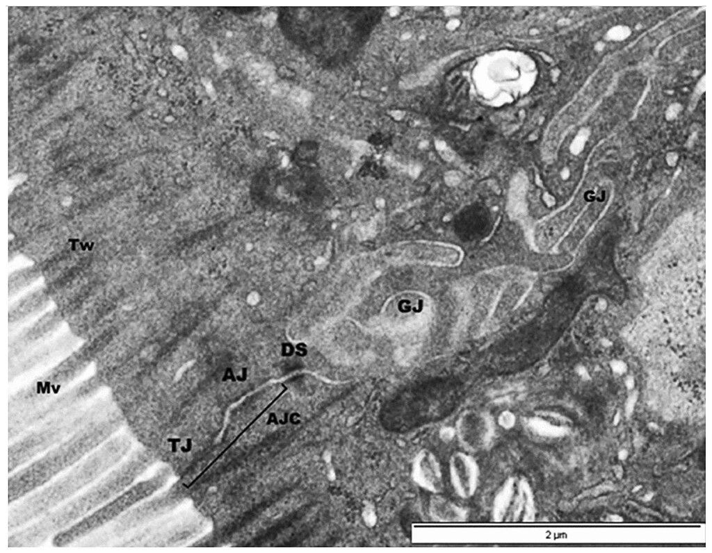
This seamless electrical communication network turns millions of individual cells into a functional syncytium. The term sounds complex, but the idea is simple. Even though the cells are structurally separate, the gap junctions make them behave as if they were one single, giant cell. When one cell gets the signal to contract, the signal flashes through the gap junctions to all its neighbors, triggering a perfectly coordinated, wave-like contraction across the entire chamber.
The Electrical Insulator
If the atria and ventricles were connected in one big syncytium, they would contract at the same time, which would be disastrously inefficient. The heart needs the atria to contract first to fill the ventricles, and then the ventricles to contract to pump blood out. To ensure this delay, the heart has an ingenious solution: the fibrous skeleton.
This is a dense framework of connective tissue, primarily collagen, that sits between the atria and the ventricles. It serves two purposes. First, it provides a rigid structure for the heart valves to attach to, ensuring they don't deform under pressure. Second, and most importantly, it acts as an electrical insulator.
The fibrous skeleton electrically isolates the atria from the ventricles, preventing the contractile signal from spreading directly between them.
This insulation forces the electrical signal to travel through a single, specific pathway: the atrioventricular (AV) node. The AV node deliberately slows the signal down for a fraction of a second before passing it to the ventricles. This slight pause is the entire reason the atria have time to finish their contraction and top up the ventricles before the ventricles begin their powerful squeeze. Without the fibrous skeleton's insulation, this critical timing would be impossible.
What is the primary function of gap junctions in cardiac muscle cells?
The heart's fibrous skeleton is crucial for proper timing of the heartbeat because it...
Understanding this micro-anatomy reveals how the heart is more than just muscle; it's a precisely engineered electrical and mechanical system, where cellular connections and insulation create the perfect rhythm of life.