Direct Digital Radiography Explained
Introduction to Radiography
Seeing the Invisible
At its heart, radiography is about making the invisible visible. It relies on a special type of light called X-rays. Like the visible light that our eyes see, X-rays are a form of electromagnetic radiation. The main difference is their energy level. X-rays have much more energy than visible light, allowing them to pass through materials that would block ordinary light, like the soft tissues of your body.
This spectrum shows where X-rays fit in. Their high energy is what gives them their unique penetrating power. Think of it like trying to throw a ball through a wall. A slowly thrown tennis ball (like visible light) will just bounce off. A speeding bullet (like an X-ray) can go right through. This simple principle is the foundation of all X-ray imaging.
Making X-rays
X-rays don't occur naturally in most environments; they have to be generated. This happens inside a device called an X-ray tube. The process is a bit like a controlled lightning strike in a vacuum.
Inside the tube, a component called a cathode is heated until it starts to release electrons. A very high voltage is then applied across the tube, which pulls these electrons toward another component, the anode, at incredible speeds. When this high-energy stream of electrons smashes into the anode, which is made of a dense metal like tungsten, the sudden stop causes the electrons to release their energy. A tiny fraction of this energy is converted into X-rays, while the rest becomes heat.
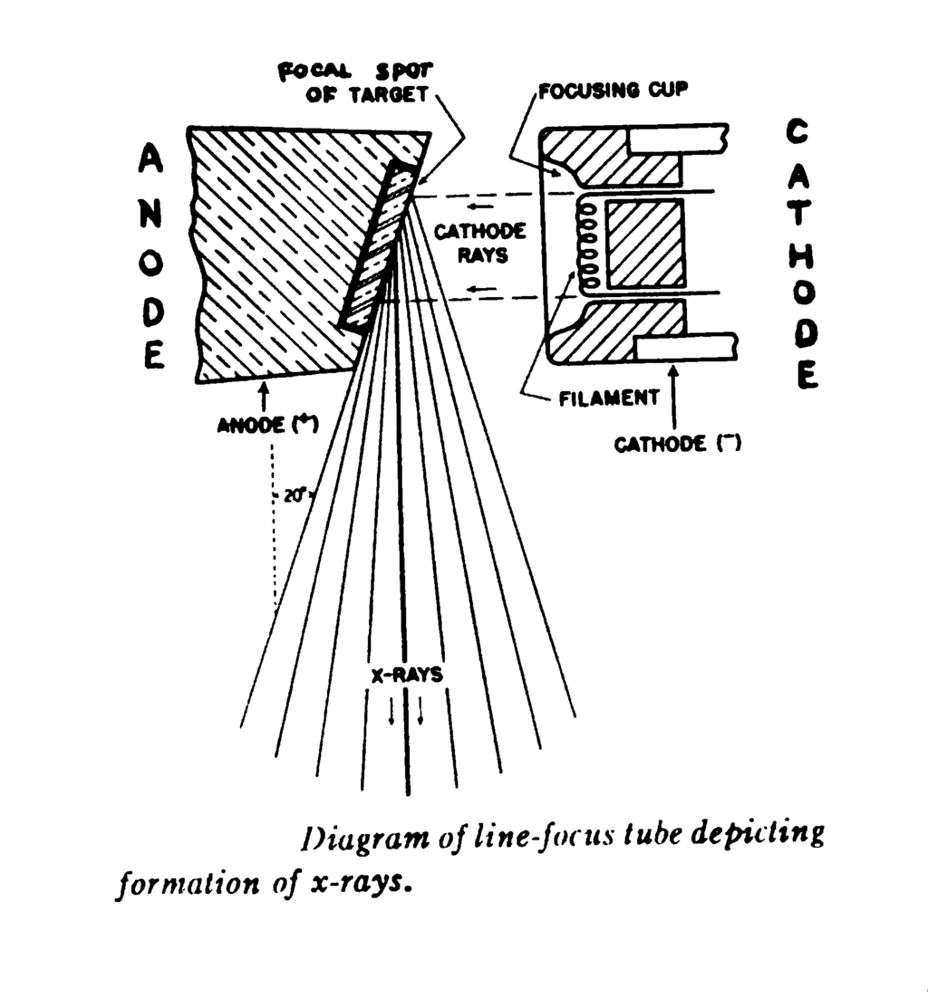
By controlling the voltage and the number of electrons, a radiographer can precisely adjust the energy and intensity of the X-ray beam for different medical procedures.
How an Image Is Formed
Once the X-rays are produced, they are directed toward the part of the body being examined. As the beam passes through the body, the different tissues interact with the X-rays in distinct ways. This interaction is primarily based on the density of the tissue.
Dense materials, like bone, contain elements with many electrons and tightly packed atoms. They are very good at absorbing or stopping X-rays. Soft tissues, like muscle and fat, are much less dense and allow most of the X-rays to pass right through. Air, such as in the lungs, is the least dense of all and barely stops any X-rays.
Dense tissue (bone) blocks many X-rays. Soft tissue (muscle) blocks some. Air blocks almost none.
On the other side of the body, a detector or a piece of photographic film records the X-rays that make it through. Areas where many X-rays are blocked, like bone, create a bright or white spot on the final image. This is because fewer X-rays reached the detector to expose it. Areas where X-rays passed through easily, like the lungs, appear dark or black because many X-rays hit the detector.
This pattern of light and dark areas creates a shadow image of the internal structures, called a radiograph.
This fundamental process of differential absorption allows doctors to see fractures in bones, identify pneumonia in the lungs, or locate foreign objects inside the body. Every X-ray image is a direct result of how different parts of you interact with this invisible, high-energy light.
Ready to check your understanding?
What is the fundamental principle that allows X-rays to create an image of internal body structures?
Inside an X-ray tube, what happens to create X-rays?
That's the basic physics behind how a simple X-ray works. By understanding how X-rays are made and how they interact with the body, we can begin to explore more advanced imaging techniques.