Diffusion Imaging in Body MRI
Introduction to MRI
How MRI Sees Inside Your Body
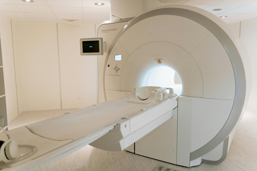
Magnetic Resonance Imaging, or MRI, is a powerful tool doctors use to see inside the human body without any surgery or radiation. It produces incredibly detailed images of organs, soft tissues, bone, and virtually all other internal body structures. But how does it work? It all starts with water and a very big magnet.
Your body is mostly water. Every water molecule () has hydrogen atoms, and at the core of each hydrogen atom is a single proton. You can think of these protons as tiny, spinning magnets. Normally, they all spin in random directions.
When you lie inside an MRI machine, you're placed in an extremely strong magnetic field—thousands of times stronger than the Earth's. This powerful field forces all those tiny proton magnets in your body to align in the same direction, much like how a compass needle points north.
The first step of an MRI is to use a powerful magnet to align the billions of protons in your body's water molecules.
Once the protons are aligned, the MRI machine sends a brief radiofrequency (RF) pulse into your body. This pulse is tuned to the right frequency to knock the spinning protons out of alignment. This is the "resonance" part of MRI. When the RF pulse is turned off, the protons start to relax back into alignment with the main magnetic field. As they do, they release the energy they absorbed from the pulse as a faint signal. The MRI's receivers detect this signal, and a computer processes it to build a detailed image.
Tissues Tell Different Stories
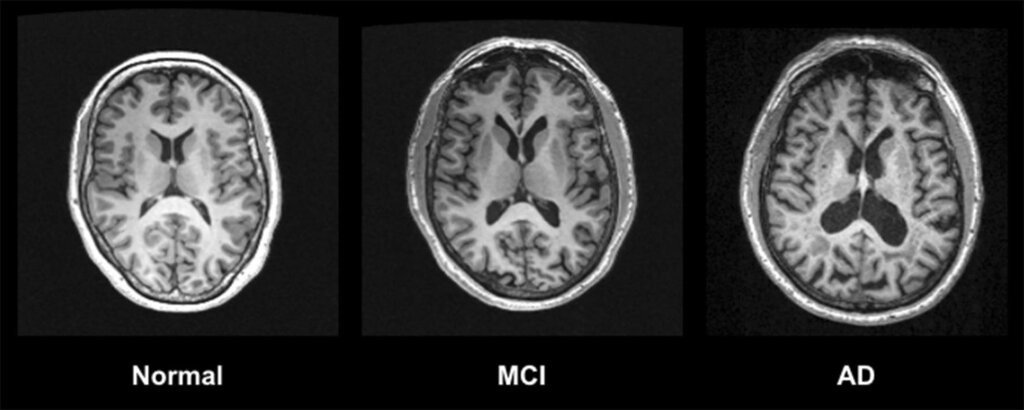
The magic of MRI is that protons in different types of body tissue relax at different speeds. By changing the timing of the RF pulses, we can highlight specific tissues. This process is called creating "weighted" images. The two most common types are T1-weighted and T2-weighted images.
T1 Relaxation
noun
The time it takes for protons to realign with the main magnetic field after the RF pulse is turned off. It's about how quickly they return to their original state.
In T1-weighted images, tissues with a short T1 time (meaning their protons realign quickly) appear bright. Fat is a great example. Tissues with a long T1 time, like water or cerebrospinal fluid (CSF), appear dark.
T2 Relaxation
noun
The time it takes for the spinning protons to lose their synchronized alignment with each other after the RF pulse. It's about how long they stay in step.
T2-weighted images are almost the opposite. Tissues where protons stay in sync for a long time (a long T2) appear bright. This makes them excellent for spotting areas of fluid or inflammation, which show up as bright signals. In T2 images, both fat and water appear bright.
| Tissue Type | T1-Weighted Image | T2-Weighted Image |
|---|---|---|
| Fat | Bright | Bright |
| Water / CSF | Dark | Bright |
| Muscle | Gray | Gray |
| White Matter | Lighter Gray | Darker Gray |
| Gray Matter | Darker Gray | Lighter Gray |
By comparing these different types of images, radiologists can identify various anatomical structures and diagnose medical conditions.
Pulse Sequences and Contrast
A pulse sequence is the specific combination and timing of radiofrequency pulses and magnetic field gradients used to create a particular type of image, like a T1 or T2-weighted scan. Technologists choose specific sequences to highlight the tissues or abnormalities they need to see.
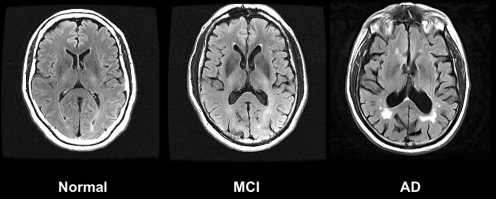
There are dozens of pulse sequences, each designed for a specific purpose. For example, a FLAIR (Fluid Attenuated Inversion Recovery) sequence is similar to a T2 image but makes the signal from cerebrospinal fluid dark. This is incredibly useful for spotting brain lesions near fluid-filled spaces.
Sometimes, doctors need even more detail. In these cases, they might use a contrast agent. The most common MRI contrast agents are based on a rare earth metal called gadolinium. When injected into the bloodstream, gadolinium shortens the T1 relaxation time of nearby protons.
This causes areas with increased blood flow—like tumors or areas of inflammation—to appear much brighter on T1-weighted images, making them stand out clearly from the surrounding healthy tissue.
Contrast agents enhance the visibility of certain structures or abnormalities by altering the relaxation times of protons in nearby tissues.
With these fundamental tools—powerful magnets, radio waves, and clever physics—MRI provides a safe and non-invasive window into the human body, helping doctors diagnose and treat a vast range of conditions.