Decoding Heart Sounds
Heart Anatomy
The Heart’s Four Rooms
Think of the heart as a duplex apartment with two units, a right side and a left side. Each unit has two rooms: an upstairs receiving area called an atrium and a downstairs pumping station called a ventricle. This gives us four chambers in total.
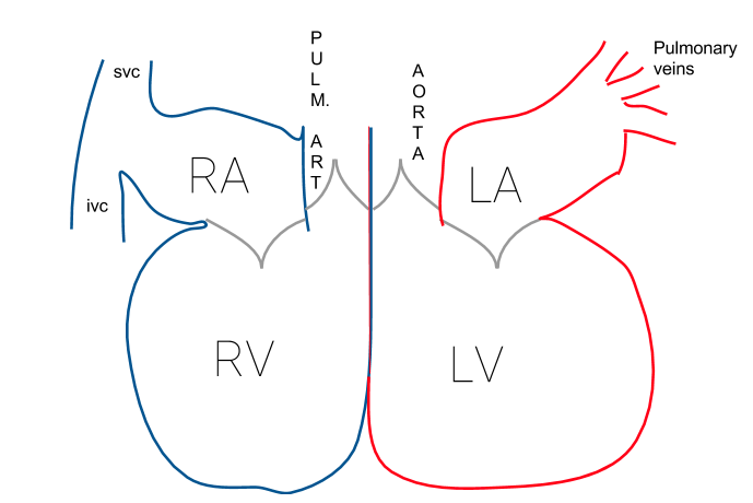
The right side of the heart handles deoxygenated blood, which is blood that has delivered its oxygen to the body's tissues and is returning for a refill.
- Right Atrium: This chamber receives the deoxygenated blood from the body.
- Right Ventricle: The right atrium pushes the blood down into this chamber, which then pumps it to the lungs to pick up fresh oxygen.
The left side of the heart deals with the newly oxygenated blood returning from the lungs.
- Left Atrium: This chamber receives the oxygen-rich blood from the lungs.
- Left Ventricle: The left atrium sends the blood down to this powerful chamber, which pumps it out to the rest of the body to deliver oxygen. The left ventricle's wall is much thicker and stronger than the right's because it has to pump blood to the entire body, not just the nearby lungs.
One-Way Doors
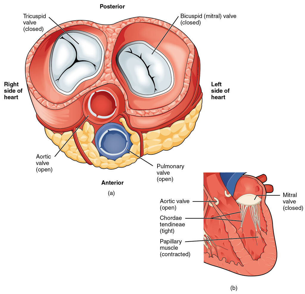
To keep blood flowing in the right direction, the heart has four valves that act like one-way doors. They open to let blood pass through and snap shut to prevent it from flowing backward. These valves are organized into two types.
The two atrioventricular (AV) valves sit between the atria and the ventricles. The two semilunar (SL) valves sit at the exits of the ventricles.
On the right side, the tricuspid valve separates the right atrium and right ventricle. On the left side, the mitral valve (also called the bicuspid valve) separates the left atrium and left ventricle.
When the ventricles pump blood out of the heart, they use the semilunar valves. The pulmonic valve is the doorway from the right ventricle to the pulmonary artery, which leads to the lungs. The aortic valve is the doorway from the left ventricle into the aorta, the body's main artery.
The Electrical Spark
The heart doesn't need the brain to tell it to beat. It has its own internal electrical system that sets the rhythm. This is the cardiac conduction system.
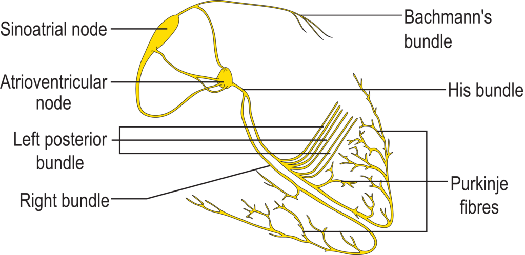
The process starts at the sinoatrial (SA) node, located in the right atrium. Often called the heart's natural pacemaker, the SA node fires off an electrical impulse that spreads across both atria, causing them to contract and push blood into the ventricles.
The signal then reaches the atrioventricular (AV) node, which sits between the atria and ventricles. The AV node acts like a gatekeeper, briefly pausing the signal. This slight delay is crucial; it gives the ventricles time to fill completely with blood before they are told to contract.
After the pause, the signal travels down a path called the bundle of His, which splits into right and left bundle branches. These branches carry the impulse down through the center wall of the heart.
Finally, the signal reaches the Purkinje fibers. These fibers branch out and spread the electrical impulse rapidly throughout the walls of both ventricles, triggering a powerful, coordinated contraction that pumps blood out to the lungs and body.
Ready to test your knowledge of the heart's anatomy?
Which chamber of the heart is the first to receive deoxygenated blood returning from the body?
Why is the wall of the left ventricle thicker and more muscular than the wall of the right ventricle?
Understanding these chambers, valves, and electrical pathways provides the foundation for figuring out how the heart produces its characteristic sounds.