Cryo-EM Fundamentals
Introduction to Cryo-EM
Seeing Life's Tiny Machines
At the heart of every living thing are molecules—tiny, intricate machines that carry out the essential tasks of life. For decades, scientists dreamed of seeing these machines in their natural state, watching them twist, turn, and interact. The challenge was immense. How do you take a picture of something thousands of times smaller than a human hair without destroying it in the process?
The answer came from a revolutionary technique called cryo-electron microscopy, or Cryo-EM. The "cryo" part refers to the extreme cold involved. Scientists flash-freeze biological samples, trapping molecules in a thin layer of glass-like ice. This process, called vitrification, is so fast that damaging ice crystals don't have time to form. The sample is preserved in a near-native state.
Once frozen, the sample is placed inside a powerful electron microscope, which uses beams of electrons instead of light to create an image. The result is a high-resolution, three-dimensional model of the molecule.
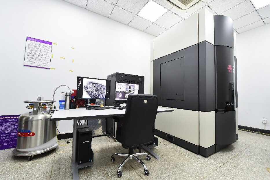
Why is this so significant? Cryo-EM allows us to see the structure of complex biological machinery that was previously invisible. We can observe how proteins fold, how viruses assemble, and how drugs bind to their targets. This opens up new frontiers in medicine, allowing for the design of more effective treatments for a wide range of diseases.
A Revolution Decades in the Making
The path to Cryo-EM was long and filled with obstacles. Early electron microscopes were harsh environments for biological samples. The intense vacuum and powerful electron beams would instantly obliterate delicate molecules.
Everything changed with a series of key breakthroughs. In the 1970s and 80s, Jacques Dubochet developed a way to vitrify water, allowing samples to be frozen without being destroyed. Around the same time, Joachim Frank created sophisticated image processing methods to combine thousands of blurry, two-dimensional images into a single, sharp three-dimensional structure. Meanwhile, Richard Henderson proved that it was possible to use an electron microscope to determine the structure of a protein at atomic resolution.
For their pioneering work, these three scientists were awarded the Nobel Prize in Chemistry in 2017. Their collective efforts transformed electron microscopy from a niche method into a mainstream tool for biological discovery.
The Structural Biologist's Toolkit
Before Cryo-EM became widely accessible, scientists primarily used two other techniques to determine the structure of molecules: X-ray crystallography and Nuclear Magnetic Resonance (NMR) spectroscopy.
X-ray crystallography has been the workhorse of structural biology for over a century. It can produce incredibly detailed atomic models, but it has a major requirement: the molecule must first be persuaded to form a highly ordered crystal. This process can be difficult, time-consuming, and sometimes impossible for large, flexible, or complex molecules. Many important biological machines simply refuse to crystallize.
NMR spectroscopy, on the other hand, studies molecules in solution, which is closer to their natural environment. However, it works best for smaller, soluble proteins and struggles to provide high-resolution data for the large molecular complexes that Cryo-EM excels at.
| Technique | How It Works | Best For | Limitations |
|---|---|---|---|
| Cryo-EM | Flash-freezes molecules and images them with electrons. | Large, complex, or flexible molecules. | Historically lower resolution than crystallography. |
| X-ray Crystallography | Bounces X-rays off a crystallized molecule. | Molecules that form stable crystals. | Requires crystallization; not ideal for flexibility. |
| NMR Spectroscopy | Measures magnetic properties of atomic nuclei. | Small to medium-sized proteins in solution. | Limited by molecular size. |
Cryo-EM filled a crucial gap. It doesn't require crystals and can handle massive molecular assemblies, from ribosomes (the cell's protein factories) to entire viruses. It allows us to see these structures in a state that's much closer to how they exist inside a cell.
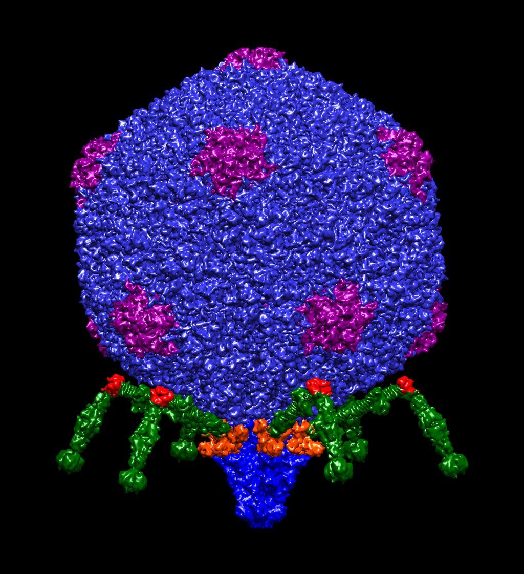
By providing this new window into the molecular world, Cryo-EM has given researchers an unprecedented ability to understand the fundamental mechanics of life.
What is the primary purpose of the 'cryo' step in cryo-electron microscopy?
Which of the following is a major advantage of Cryo-EM compared to X-ray crystallography?