Computed Tomography Explained
X-ray Fundamentals
The Invisible Light
X-rays are a form of high-energy light, invisible to our eyes. Think of the spectrum of light like a piano keyboard. On one end are low-energy radio waves, and on the other are high-energy gamma rays. Visible light sits somewhere in the middle. X-rays are much further up the keyboard, with shorter wavelengths and more energy than visible light or even ultraviolet light.
This high energy is their superpower. It allows them to pass through materials that block visible light, like skin and muscle. This simple property revolutionized our ability to see inside the human body without making a single cut.
Making X-rays
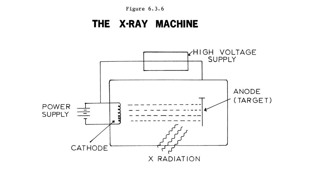
Creating X-rays happens inside a device called an X-ray tube. It’s a vacuum-sealed tube containing two key components: a cathode and an anode.
First, the cathode's filament is heated, causing it to release a cloud of electrons. Then, a high voltage is applied across the tube, creating a powerful electrical field. This field catapults the electrons from the cathode toward the anode, a metal target, at incredibly high speeds.
When these speeding electrons slam into the anode, they stop abruptly. Their immense kinetic energy has to go somewhere. Most of it converts into heat, but a small fraction transforms into X-ray photons. These newly created X-rays exit the tube through a window, ready to be used for imaging.
Creating an Image
So how do these X-rays create the images we're familiar with? It all comes down to how different materials in the body interact with the X-ray beam. As X-rays travel through the body, they are absorbed or scattered to varying degrees. This process is called attenuation.
Attenuation
noun
The reduction in the intensity of an X-ray beam as it passes through matter.
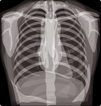
The amount of attenuation depends on the density and atomic number of the tissue. Dense structures with high atomic numbers, like bone (rich in calcium), are excellent at blocking X-rays. They cause high attenuation. Softer tissues, like muscle, fat, and organs, are less dense and let more X-rays pass through, causing lower attenuation. Air-filled spaces, like the lungs, hardly block X-rays at all.
After passing through the body, the remaining X-rays hit a detector (which used to be film, but is now usually digital). Areas on the detector that are struck by many X-rays turn dark, while areas struck by few or no X-rays remain bright. The result is a shadow image: bones appear white, soft tissues appear in shades of gray, and air appears black.
A Brief History
X-rays were discovered by accident in 1895 by German physicist Wilhelm Röntgen. He was studying cathode rays when he noticed a fluorescent screen in his lab glowing, even though it was shielded from the cathode ray tube. He realized he had stumbled upon a new, unknown type of ray, which he called "X-rays," with 'X' signifying the unknown.
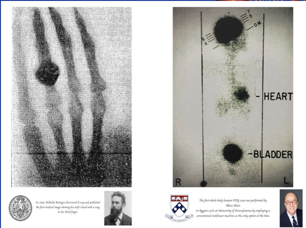
Within weeks of his discovery, Röntgen took the first medical X-ray image: a picture of his wife's hand, clearly showing her bones and wedding ring. The medical potential was immediately obvious. This groundbreaking discovery earned him the first-ever Nobel Prize in Physics in 1901 and laid the foundation for diagnostic radiology.
Let's review the key concepts before moving on.