Clinical Medical Terminology Applications
Cardiovascular Pathophysiology
When Valves Go Wrong
The heart's four valves act as one-way doors, ensuring blood flows in the right direction. When they function correctly, they open and close with perfect timing. But disease can damage these delicate structures, leading to two main problems: stenosis and regurgitation.
Stenosis
noun
The narrowing or stiffening of a heart valve, which restricts blood flow out of a chamber. The heart must work harder to pump blood through the smaller opening.
Imagine trying to push a crowd through a door that's only halfway open. That's stenosis. The valve leaflets become stiff, often from calcium buildup or scarring, and can't open fully. This creates a bottleneck, forcing the heart chamber behind it to generate much higher pressure to eject blood.
Regurgitation
noun
A condition where a heart valve doesn't close tightly, allowing blood to leak backward. This is also known as insufficiency or incompetence.
Regurgitation, on the other hand, is like a door that doesn't latch. The valve fails to seal properly, so with each heartbeat, some blood flows backward. This backflow, or regurgitant jet, means the heart has to pump the same blood twice, increasing its workload and causing volume overload in the chamber receiving the leaked blood.
The Aftermath of a Heart Attack
A heart attack, or myocardial infarction, occurs when blood flow to a part of the heart muscle is blocked, causing cells to die. The immediate event is dangerous, but the long-term structural changes, known as remodeling, create their own set of problems. The heart muscle, now weakened and scarred, can stretch and change shape.
This leads to a crucial distinction in medical charting. Cardiomegaly simply means an enlarged heart, which can be seen on an X-ray. It's a sign, not a diagnosis. Cardiomyopathy, however, is a disease of the heart muscle itself that makes it harder for the heart to pump blood. The enlargement (cardiomegaly) is often a result of the underlying muscle disease (cardiomyopathy).
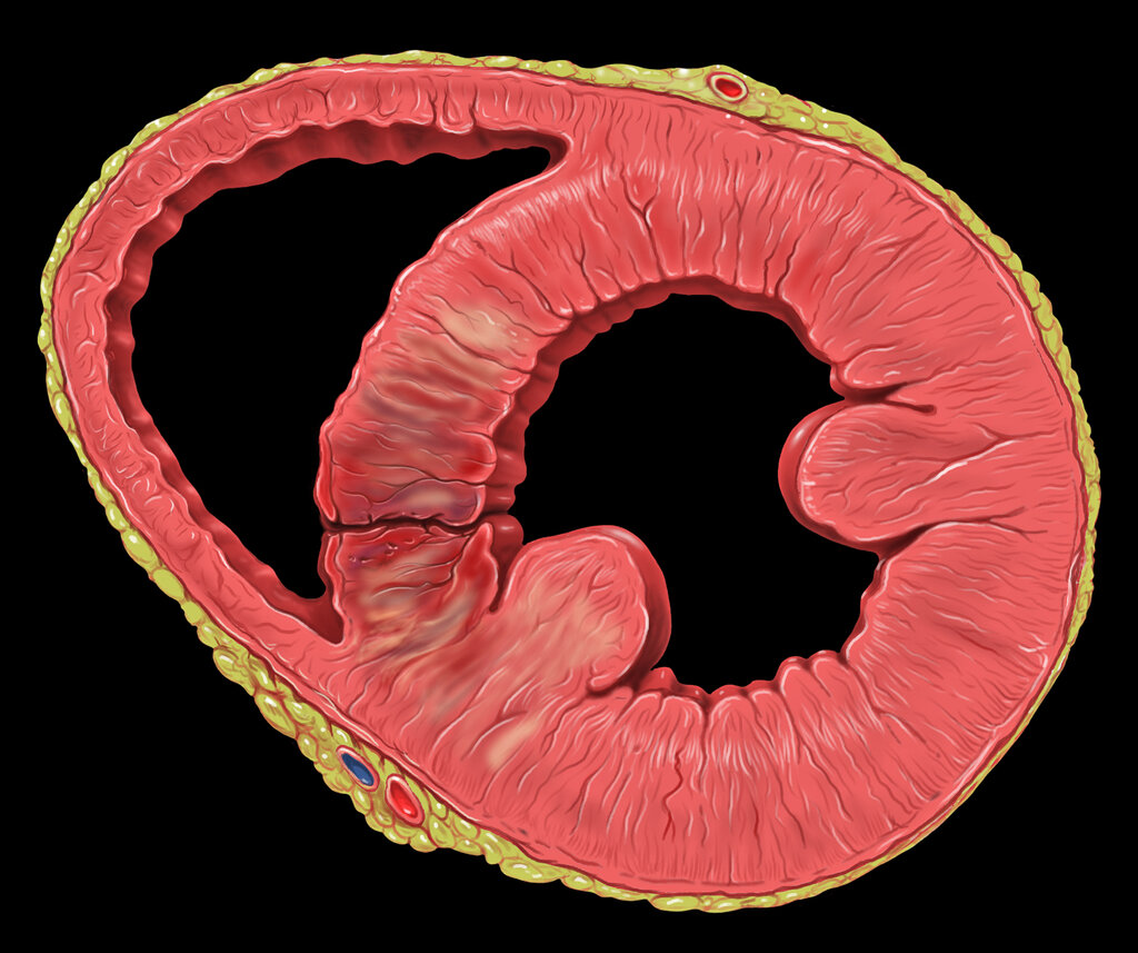
Beyond general weakening, specific mechanical complications can arise. If the inner wall of the ventricle is damaged, a mural thrombus (a blood clot attached to the wall) can form, posing a risk of traveling to the brain and causing a stroke. In severe cases, the dead tissue can rupture days after the initial attack. A rupture of the ventricular free wall leads to a rapid, often fatal condition called cardiac tamponade. This happens when blood floods the pericardial sac, the space around the heart, compressing it and preventing it from filling properly.
Disruptions in Rhythm
The heart's electrical conduction system is a marvel of biological engineering, coordinating the heartbeat with precise signals. When this system malfunctions, it causes an arrhythmia, an irregular heartbeat. Arrhythmias are broadly classified based on their speed (too fast or too slow) and their origin.
Tachycardias are fast heart rhythms ( beats per minute), while bradycardias are slow rhythms ( beats per minute). They are further divided into supraventricular (originating above the ventricles, in the atria or AV node) and ventricular (originating within the ventricles). Ventricular arrhythmias are generally more dangerous because they can severely compromise the heart's ability to pump blood.
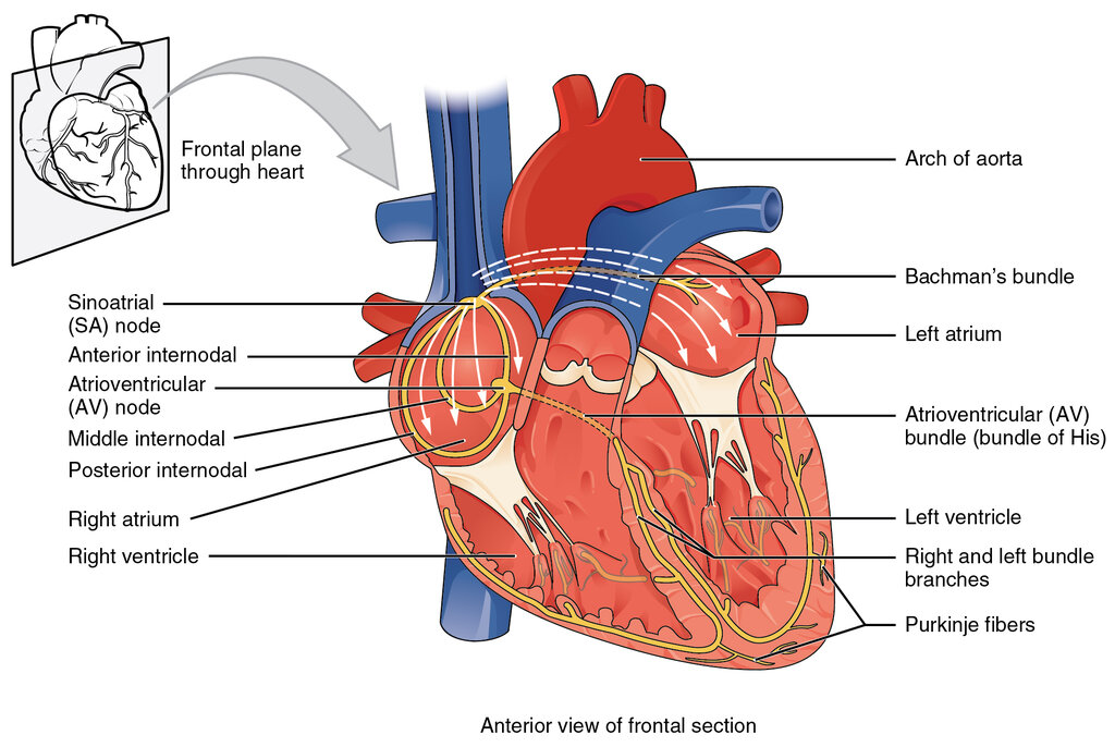
For example, (A-fib) is a common supraventricular arrhythmia where the atria quiver chaotically instead of contracting effectively. While not immediately life-threatening, it reduces cardiac efficiency and significantly increases the risk of stroke due to blood pooling in the atria. In contrast, ventricular tachycardia is a rapid rhythm from the ventricles that can quickly degenerate into ventricular fibrillation, a state of chaotic quivering that causes sudden cardiac arrest.
Pressure, Flow, and Resistance
Understanding heart failure requires a grasp of hemodynamics, the physics of blood flow. Key terms describe the forces the heart works with and against. Preload is the stretch on the ventricular muscle at the end of diastole (the filling phase). Think of it as the volume of blood loading up the ventricle before it contracts. Afterload is the resistance the ventricle must overcome to eject blood. It's largely determined by blood pressure in the aorta and the systemic circulation.
These terms are crucial for understanding pathology. In heart failure, for example, a weakened ventricle may not be able to handle a normal preload, leading to a backup of fluid in the lungs or body. Medications often target these forces, aiming to reduce preload or afterload to ease the heart's workload.
Cardiac Output () is the total volume of blood pumped by the heart per minute. It's the ultimate measure of heart function.
Finally, it's important to clarify the difference between two often-confused terms related to blood vessels. Arteriosclerosis is a general term for the hardening and loss of elasticity of arteries, a natural part of aging. is a specific type of arteriosclerosis caused by the buildup of fatty plaques within the artery walls. This plaque buildup is the primary culprit behind most heart attacks and strokes.
With these terms, a patient's chart becomes a narrative. A diagnosis of 'severe aortic stenosis with concentric left ventricular hypertrophy' tells a story of a narrowed valve forcing the heart muscle to thicken and work dangerously hard. This language is the bridge between anatomy and the living reality of heart disease.
Now, let's test your understanding of how these concepts apply in clinical scenarios.
A patient's echocardiogram shows that their mitral valve fails to seal properly, causing some blood to flow backward with each heartbeat. What is the correct term for this condition?
Which of the following describes the resistance the heart must overcome to eject blood into the aorta?
Understanding how structural problems create functional disease is the core of pathophysiology. It moves beyond simple identification of parts to a dynamic understanding of the heart as a system under pressure.