Clinical Mastery of Microneedling and Fractional Lasers
Cellular Wound Response
The Controlled Cascade
When a microneedling device creates a micro-injury, the body doesn't perceive it as a major wound. Instead, it initiates a precise and localized healing cascade, a beautifully orchestrated series of cellular events. This process begins within seconds as platelets rush to the site. They aren't just there to clot; they are messengers, releasing a cocktail of growth factors, chief among them being platelet-derived growth factor (PDGF).
Microneedling, also known as collagen induction therapy, is a cutting-edge technique of designed to rejuvenate and revitalize the skin.
PDGF signaling acts like a foreman at a construction site, shouting orders to the primary cellular workforce. It sends a powerful chemotactic signal that calls fibroblasts to migrate into the area. Simultaneously, it triggers the release of other critical signals, including Vascular Endothelial Growth Factor (VEGF) and Fibroblast Growth Factor (FGF). Unlike laser treatments that create a zone of thermal damage, this stimulation is purely mechanical, preserving the surrounding tissue and allowing for a more controlled regenerative response.
Building and Remodeling
Once fibroblasts arrive, they begin the process of neocollagenesis. This phase is heavily modulated by the Transforming Growth Factor-beta (TGF-β) family of proteins. Initially, the fibroblasts lay down a scaffold of fine, disorganized Type III collagen. This is the body's quick-fix material, the cellular equivalent of spackle. It provides structure but lacks the tensile strength of mature skin.
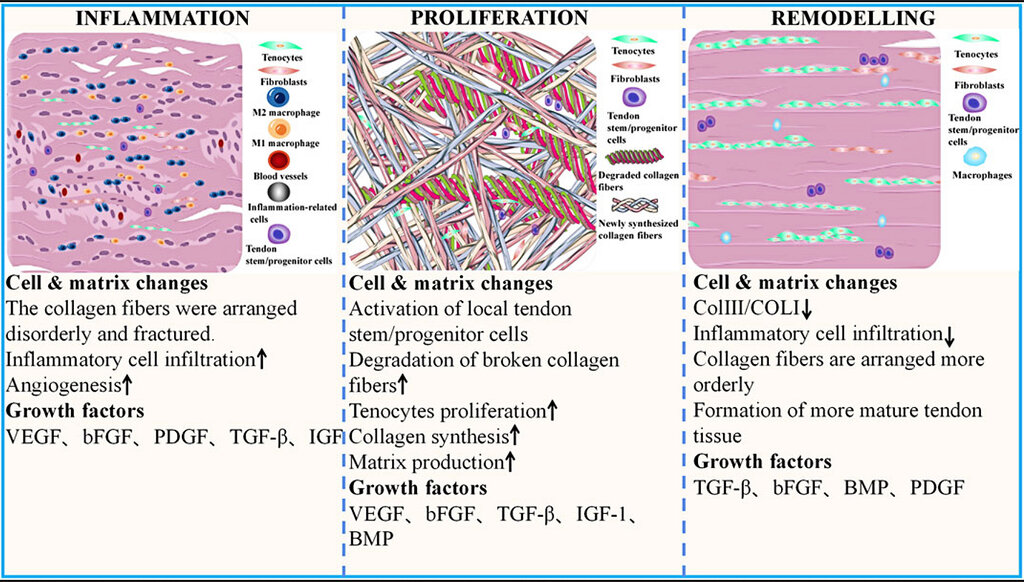
The specific isoforms of TGF-β play distinct roles. TGF-β1 and TGF-β2 are potent stimulators of collagen production but are also implicated in fibrosis and scar formation. The magic of CIT lies in its ability to preferentially stimulate TGF-β3, which promotes the synthesis of new collagen while simultaneously inhibiting the fibrotic pathways. This isoform encourages a more organized, basket-weave deposition of collagen, mimicking healthy tissue rather than scar tissue. At the same time, the release of VEGF and FGF stimulates neoangiogenesis, the formation of new capillaries that supply the regenerating tissue with oxygen and nutrients.
The Final Architecture
The initial wound-filling phase gives way to a longer remodeling period that can last for months. During this time, the cellular construction crew begins to refine the structure. Enzymes called (MMPs) are activated. Their job is to break down the temporary Type III collagen scaffold. As they clear away the old material, fibroblasts replace it with robust, highly organized Type I collagen, the primary structural protein in the dermis. This carefully managed transition from Type III to Type I collagen is what ultimately leads to increased skin firmness and elasticity.
This response also involves intricate communication between other cell types. After the injury, keratinocytes at the site upregulate signaling molecules. This communication can influence melanocytes, the pigment-producing cells. In a well-controlled healing environment like that from CIT, this cross-talk is managed, minimizing the risk of post-inflammatory hyperpigmentation that can occur with more aggressive or thermally damaging treatments.
Ultimately, Collagen Induction Therapy leverages the body's own sophisticated, multi-stage repair mechanisms. It initiates a controlled inflammatory and proliferative response that culminates in a long-term remodeling phase, resulting in a denser, healthier, and more organized dermal matrix.