Clinical Mastery of Cyanotic and Acyanotic Heart Disease
Hemodynamics of Shunting
Shunts and Resistance
In a healthy circulatory system, the pulmonary and systemic circuits are separate. Blood flows in a single, continuous loop. But a congenital heart defect can create a hole or connection between these two circuits, causing blood to take a shortcut. This abnormal flow is called a shunt.
The direction and volume of a shunt are not random. They are governed by the path of least resistance. Two key forces are at play: systemic vascular resistance (SVR) and pulmonary vascular resistance (PVR). SVR is the resistance the left ventricle must overcome to pump blood to the entire body. PVR is the resistance the right ventricle faces when pumping blood to the lungs.
Before birth, a fetus's lungs are collapsed and filled with fluid, making PVR extremely high. The placenta provides a low-resistance circuit, so SVR is low. After a baby's first breath, the lungs expand, and PVR plummets dramatically. Simultaneously, the placental connection is lost, causing SVR to rise. This switch is fundamental to understanding why shunts behave the way they do.
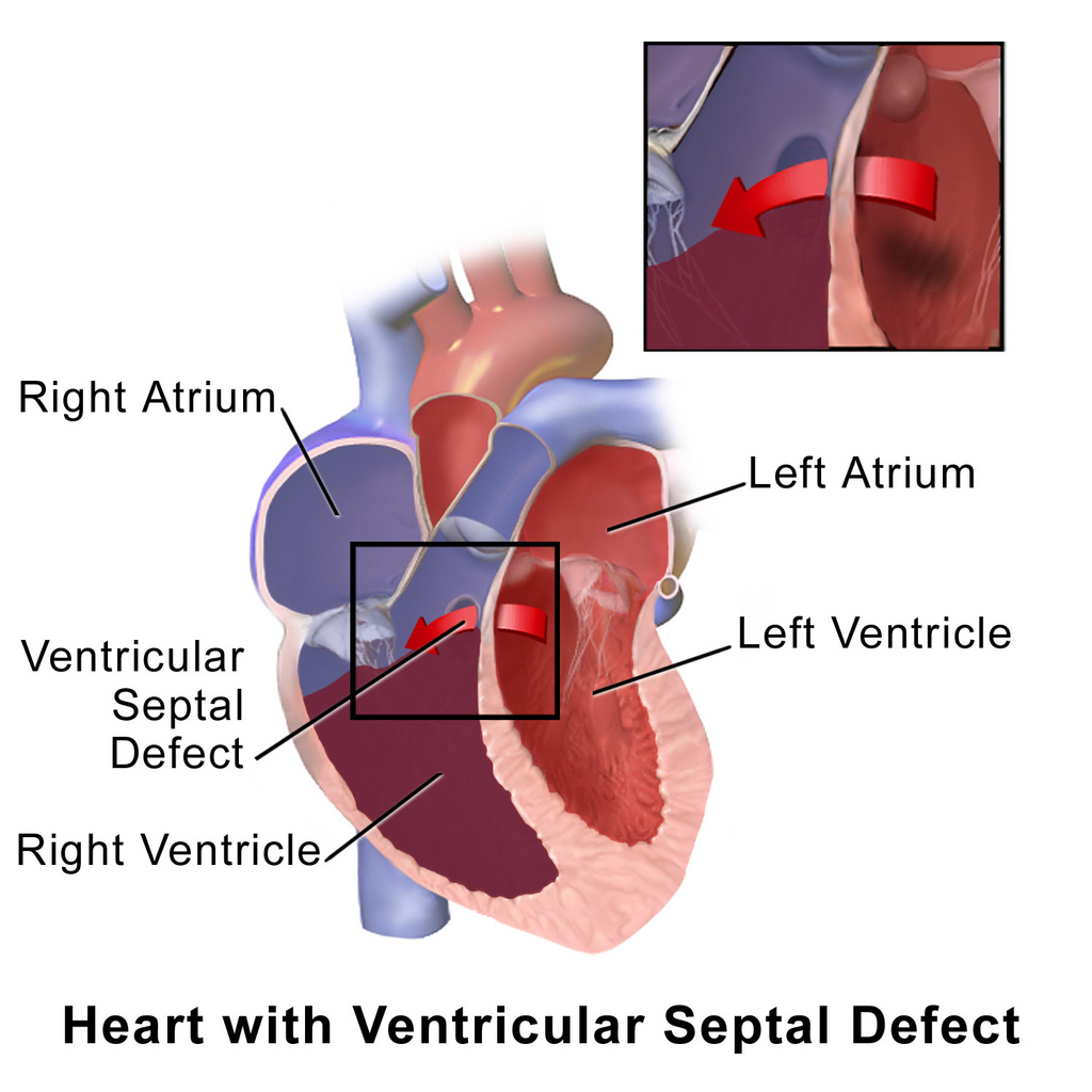
Postnatal circulation is a high-pressure system on the left and a low-pressure system on the right. When a defect like an atrial septal defect (ASD), ventricular septal defect (VSD), or patent ductus arteriosus (PDA) is present, blood naturally flows from the high-pressure left side to the lower-pressure right side. This is called a left-to-right shunt.
This type of shunt results in an excessive volume of blood being pumped to the lungs. Because this shunted blood is already oxygenated, the patient does not typically appear blue. This is why these conditions are known as acyanotic heart defects. However, the increased pulmonary blood flow puts a significant strain on the right ventricle and the lung's blood vessels.
Quantifying the Shunt
To determine if a shunt is severe enough to warrant intervention, cardiologists measure the ratio of pulmonary blood flow (Qp) to systemic blood flow (Qs). In a normal heart, the amount of blood going to the lungs is equal to the amount going to the body, so the Qp:Qs ratio is 1:1.
With a left-to-right shunt, Qp becomes greater than Qs. This ratio can be calculated using oxygen saturation levels measured during a cardiac catheterization, based on the Fick principle. The calculation compares the amount of oxygen picked up in the lungs to the amount consumed by the body.
A Qp:Qs ratio greater than 1.5:1 is generally considered hemodynamically significant. This means the lungs are receiving at least 50% more blood flow than the rest of the body. Such a large, sustained volume overload can lead to irreversible damage to the pulmonary blood vessels, a condition called pulmonary hypertension. Therefore, a ratio above this threshold is a primary driver for considering surgical or catheter-based closure of the defect.
With such defect, the high pressure from the left side shunts blood from left to right – which has lower pressure; the pulmonary vascular resistance (PVR) is less than the systemic vascular resistance (SVR) and this allows for more blood to shunt to the lungs, overwhelming the lungs, and therefore causing pulmonary edema and decreased lung compliance.
Flow Reversal and Classification
What happens if the pressure on the right side of the heart exceeds the left? The shunt reverses, becoming a right-to-left shunt. In this scenario, deoxygenated blood from the right side bypasses the lungs and flows directly into the systemic circulation. This lowers the overall oxygen content of the blood, leading to cyanosis, or a bluish discoloration of the skin.
This reversal can happen for two main reasons. First, a structural defect may cause a severe obstruction to blood flow into the pulmonary artery, raising right-sided pressures. Second, a long-standing left-to-right shunt can cause so much damage that the PVR becomes permanently fixed at a very high level, eventually exceeding SVR. This severe, irreversible pulmonary hypertension is known as —a late and serious complication.
The size of the defect and the compliance of the heart's chambers also influence the shunt's magnitude. A large, non-restrictive VSD allows pressures between the ventricles to equalize, so the shunt is determined almost entirely by the PVR/SVR ratio. A small, restrictive defect limits flow, so a large pressure difference can exist without a large shunt.
Furthermore, the stiffness or compliance of the ventricles affects diastolic filling. If the left ventricle is stiff (poor compliance), it can raise left atrial pressure during diastole, increasing a left-to-right shunt through an ASD even if systolic pressures are normal.
To account for these complexities, the 2025 ACC/AHA updated classification system for CHD integrates both anatomy and physiology. This Anatomic-Physiological (AP) system provides a more nuanced way to categorize defects based not just on what they are, but on how they affect blood flow, pressure, and oxygen levels, guiding more precise clinical decision-making.
What is the primary factor that determines the direction of blood flow through a cardiac shunt?
Immediately after a baby takes its first breath, which of the following circulatory changes occurs?
Understanding the interplay of pressure, resistance, and flow is key to managing congenital heart disease. It explains not only why shunts occur but also how to quantify their impact and decide when to intervene.