Clinical Management of Lead Poisoning
Pathophysiology and Toxicokinetics
Lead's Cellular Deception
Lead is a master of disguise. Its toxicity stems from its ability to mimic essential metals that our bodies rely on, particularly calcium, zinc, and iron. Because of its similar ionic radius and charge, lead can sneak into cellular pathways designed for these vital elements, disrupting fundamental processes. This chemical impersonation is the root of its widespread damage.
Nowhere is this more apparent than with calcium. Lead competes with calcium at binding sites, interfering with nerve impulse transmission, muscle contraction, and the function of countless enzymes. It hijacks the very signalling systems that calcium ions () regulate. By displacing calcium, lead can disrupt the blood-brain barrier, alter neurotransmitter release, and trigger apoptosis, or programmed cell death.
A Long and Damaging Journey
Once absorbed, lead doesn't stay in one place. Its movement through the body is best described by a three-compartment model, which divides its distribution into blood, soft tissues, and mineralised tissue (bones and teeth).
Initially, lead enters the blood, where it has a half-life of about 30 days. From there, it distributes to soft tissues like the liver, kidneys, and brain. Over time, however, the vast majority—up to 95% in adults—is sequestered into bone, where it takes the place of calcium in the bone matrix. In bone, lead is far more stable, with a half-life measured in decades.
This long-term storage creates a dangerous internal reservoir. During periods of bone turnover, such as pregnancy, menopause, or when a fracture heals, this stored lead can be remobilised back into the bloodstream, causing a recurrence of toxic effects years after the initial exposure has ended. This also explains the 'rebound' effect seen after , where blood lead levels drop during treatment but then rise again as lead leaches out from bone stores.
The Heme Synthesis Saboteur
One of lead's most significant toxic effects is its disruption of heme synthesis. Heme is a critical component of haemoglobin, the protein in red blood cells that carries oxygen. Lead interferes with this multi-step process by inhibiting key enzymes.
Lead specifically inhibits two enzymes in the heme production line: aminolevulinic acid dehydratase (ALAD) and ferrochelatase.
The inhibition of causes a buildup of its substrate, aminolevulinic acid (ALA), which is itself neurotoxic. Measuring ALA in the urine is a common biomarker for lead poisoning.
Further down the pathway, lead blocks , the final enzyme in the chain. Ferrochelatase is responsible for inserting iron into the heme molecule. When this step is blocked, a zinc-containing molecule (zinc protoporphyrin) is formed instead of heme. The resulting lack of functional haemoglobin contributes to anaemia, a classic sign of chronic lead toxicity.
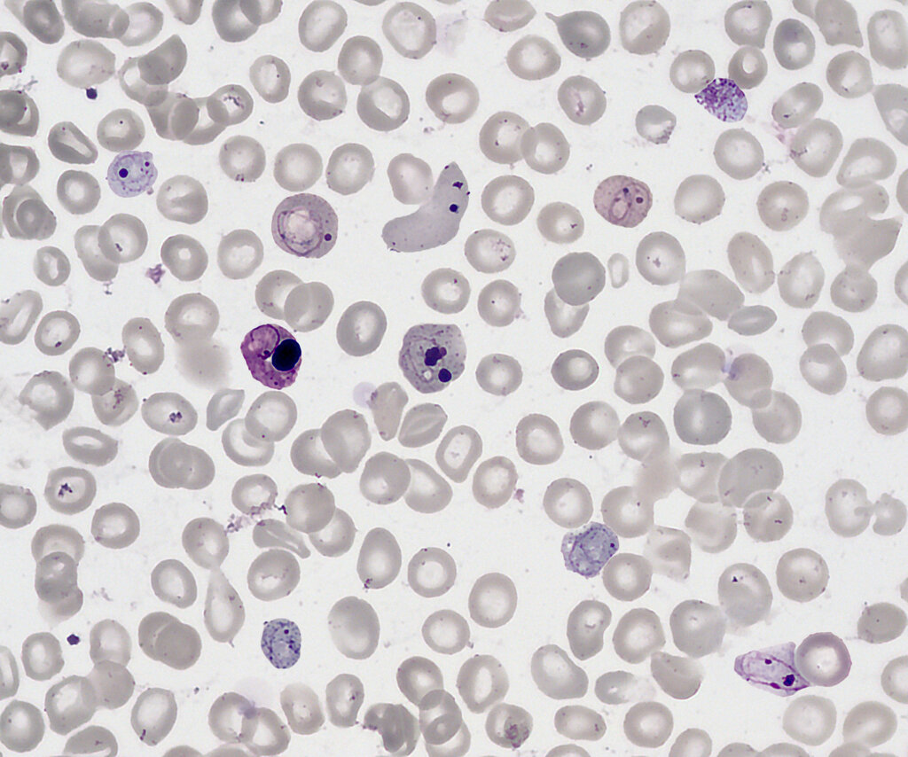
This dual-pronged attack on heme synthesis cripples the body's ability to produce one of its most essential molecules, with cascading effects on oxygen transport and cellular energy.
Lead's toxicity primarily stems from its ability to chemically mimic which group of essential metals?
According to the three-compartment model, where is the vast majority (up to 95%) of lead stored long-term in the adult body?