Clinical Management of Gastric Cancer
Staging and Histopathology
Decoding Gastric Cancer
Not all gastric cancers are the same. To treat the disease effectively, doctors first need to understand its specific characteristics. This process of classification looks at the cancer's appearance under a microscope, its physical location and spread, and its underlying genetic makeup. These details guide everything from prognosis to treatment strategy.
Classification is the language doctors use to describe a specific cancer's behaviour and to plan the best attack.
The Lauren Classification
One of the most enduring systems for classifying gastric adenocarcinoma is the Lauren classification, proposed in 1965. It divides tumours into two main types based on their growth patterns: intestinal and diffuse.
The Intestinal type is characterised by cohesive cells that group together to form gland-like structures, similar to those found in the intestine. These tumours are often associated with chronic inflammation, such as that caused by H. pylori infection, and tend to be more common in older males. Because they form distinct masses, they are often more readily detected during endoscopy.
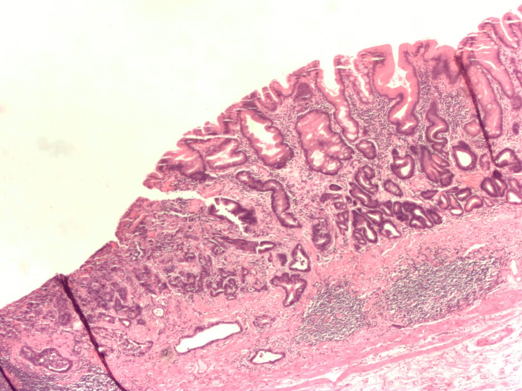
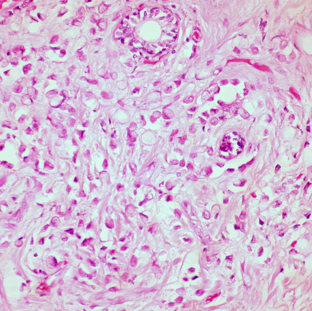
The Diffuse type, in contrast, is more insidious. Its cells are non-cohesive and infiltrate the stomach wall individually or in small clusters, thickening the wall without forming a discrete mass. This type is more common in younger patients and is notorious for the presence of signet ring cells—cells where mucus pushes the nucleus to the periphery. Diffuse cancers often have a poorer prognosis due to their infiltrative nature and tendency to be diagnosed at a later stage.
A third category, Mixed type, exhibits features of both intestinal and diffuse cancers. The World Health Organisation (WHO) provides a more detailed breakdown of histological subtypes, but the Lauren system remains a clinically useful shorthand.
| WHO Histological Type | Key Features |
|---|---|
| Papillary Adenocarcinoma | Finger-like projections |
| Tubular Adenocarcinoma | Branching, tube-like glands |
| Mucinous Adenocarcinoma | >50% of the tumour is extracellular mucin |
| Poorly Cohesive Carcinoma | Includes signet ring cell type; diffuse infiltration |
| Mixed Adenocarcinoma | A mixture of different types |
Staging with TNM
Once the histology is known, the cancer is staged to determine its extent. The universally accepted system is the AJCC (American Joint Committee on Cancer) TNM staging system, now in its 8th Edition. It assesses three key factors:
- T (Tumour): How deeply has the primary tumour invaded the stomach wall?
- N (Nodes): Has the cancer spread to nearby lymph nodes, and if so, how many?
- M (Metastasis): Has the cancer spread to distant parts of the body?
One common system is TNM, which assigns a number to the size, location and growth of the tumor (T); whether it has spread to the lymph nodes (N); and how far the cancer has spread from its original location, or metastasized (M).
The 8th edition brought important changes, particularly for tumours at the gastroesophageal junction (GEJ). These are classified using the , which divides them based on the location of the epicentre of the tumour relative to the junction. A key change in the 8th edition is that Siewert type III tumours are now staged as esophageal cancers, not gastric cancers, reflecting their distinct biological behaviour.
The N (node) classification also became more granular. The N3 category was split into N3a (7 to 15 positive lymph nodes) and N3b (16 or more positive nodes). This distinction acknowledges that a higher burden of lymph node involvement corresponds to a significantly worse prognosis.
Molecular Subtypes
Beyond what can be seen under a microscope, the genetic and molecular profile of a tumour offers powerful insights. Large-scale projects like The Cancer Genome Atlas (TCGA) have identified four major molecular subtypes of gastric cancer, each with unique features and potential therapeutic targets.
| Subtype | Frequency | Key Features | Potential Therapies |
|---|---|---|---|
| EBV (Epstein-Barr Virus) | ~9% | Associated with EBV infection; extreme DNA hypermethylation; high expression of PD-L1/L2. | Immunotherapy (checkpoint inhibitors) |
| MSI (Microsatellite Instability) | ~22% | High mutation rate; older age of onset; often intestinal type. | Immunotherapy (checkpoint inhibitors) |
| GS (Genomically Stable) | ~20% | Low mutation rate; often diffuse histology; younger age of onset. | Difficult to target; chemotherapy. |
| CIN (Chromosomal Instability) | ~50% | Widespread DNA copy number alterations; often intestinal type; common at the GEJ. | Targeted agents (e.g., HER2 inhibitors) |
This molecular classification is revolutionising treatment. For instance, tumours that are positive for EBV or have are often highly responsive to immunotherapy drugs called checkpoint inhibitors. Similarly, about 20% of CIN tumours overexpress a protein called HER2, making them candidates for targeted drugs like trastuzumab. Understanding these subtypes allows for a more personalised and effective approach to treating gastric cancer.
By combining histological, anatomical, and molecular data, oncologists can build a comprehensive profile of a patient's cancer, leading to more accurate prognoses and tailored treatment plans.
According to the Lauren classification, what is a key characteristic of diffuse-type gastric cancer?
What significant change regarding gastroesophageal junction (GEJ) tumours was introduced in the 8th Edition of the AJCC TNM staging system?