Clinical Management of Femoral Hernias
Femoral Canal Boundaries
The Femoral Triangle
To understand the femoral canal, we first need to look at its location: the femoral triangle. This is a wedge-shaped area in the upper thigh. Its borders are formed by three structures:
- Superiorly (top): The inguinal ligament.
- Laterally (outside): The sartorius muscle.
- Medially (inside): The adductor longus muscle.
Within this triangle lie several critical structures that pass between the torso and the leg.
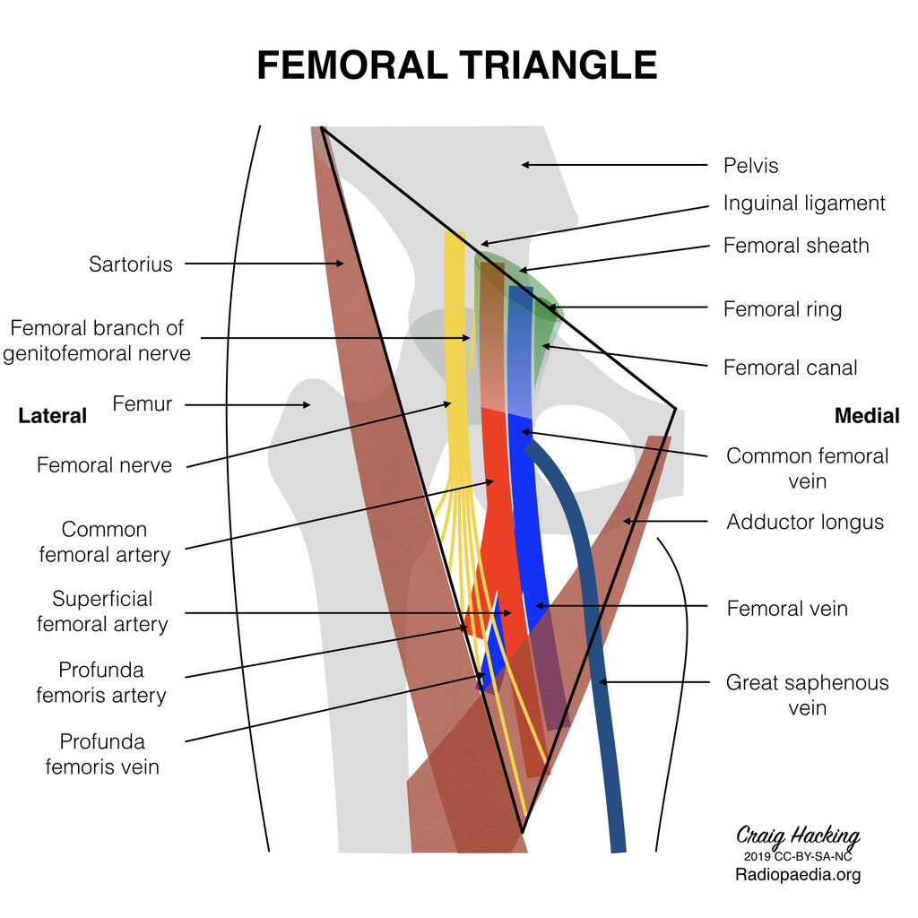
Inside the femoral triangle is the femoral sheath, a funnel-shaped sleeve of fascia. This sheath doesn't contain the femoral nerve, but it does wrap around the femoral artery, femoral vein, and the femoral canal. It's divided into three compartments.
The structures of the femoral triangle, from lateral to medial, can be remembered as, NAVEL
This mnemonic stands for:
- Nerve (Femoral)
- Artery (Femoral)
- Vein (Femoral)
- Empty Space (The Femoral Canal)
- Lymphatics (in the Femoral Canal)
The femoral nerve sits outside the femoral sheath, but the artery, vein, and canal are neatly packaged within it.
The Femoral Ring and Canal
The femoral canal is the smallest, most medial compartment of the femoral sheath. It's only about 1.25 cm long and serves as a passageway for lymphatic vessels. At its base is a lymph node known as the deep inguinal lymph node of Cloquet. While it seems like just empty space, this canal is a critical point of weakness in the abdominal wall.
The entrance to this canal is called the femoral ring. This ring is bordered by structures that are rigid and unforgiving, which is key to its clinical importance.
| Boundary | Structure |
|---|---|
| Anterior | Inguinal ligament |
| Posterior | Pectineal ligament |
| Medial | Lacunar ligament |
| Lateral | Femoral vein |
Because these boundaries—two ligaments, a vein, and bone (via the pectineal ligament)—don't stretch, anything that pushes through the femoral ring is at high risk of getting trapped. When abdominal contents, like a loop of intestine, protrude through this tight opening, it's called a femoral hernia. The unyielding nature of the ring means the herniated tissue can easily have its blood supply cut off, a dangerous condition known as strangulation.
The image below shows a CT scan of a patient with a femoral hernia. You can see the small bowel loop has passed through the femoral canal.
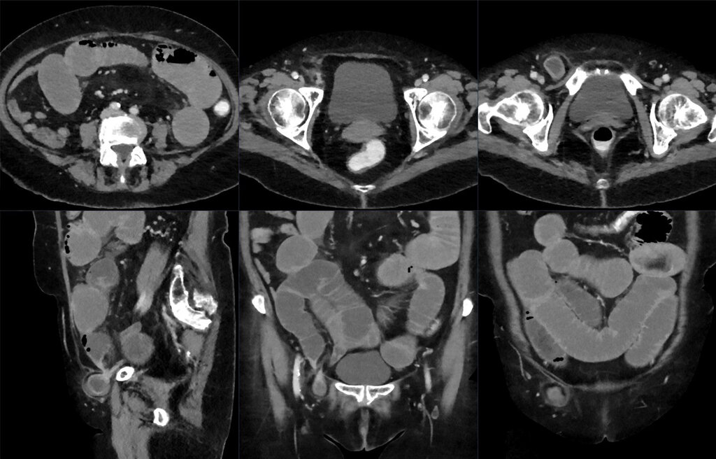
Understanding the rigid anatomy of the femoral ring is essential. It directly explains why femoral hernias, though less common than inguinal hernias, are considered more of a surgical emergency due to the high risk of incarceration.
Which of the following structures is NOT a border of the femoral triangle?
Which of the following structures is located within the femoral sheath?