Clinical Management of Femoral Hernias
Femoral Canal Anatomy
The Femoral Sheath and Canal
Deep within the groin, where the thigh meets the abdomen, lies a critical anatomical region. The major blood vessels supplying the lower limb pass through here, protected by a fascial sleeve called the femoral sheath. Think of this sheath as a funnel, extending downwards from the abdomen into the upper thigh. It doesn't contain the femoral nerve, which passes into the thigh just lateral to it, but it does house the primary femoral vessels.
The femoral sheath is divided by thin walls, or septa, into three distinct compartments. From lateral to medial, they are:
- Lateral Compartment: Contains the femoral artery.
- Intermediate Compartment: Contains the femoral vein.
- Medial Compartment: This is the femoral canal.
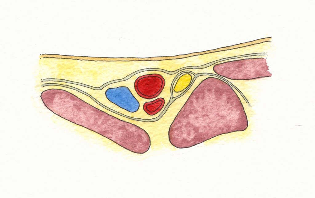
The femoral canal is the smallest of the three compartments. It's a short, conical space, typically only about 1.25 cm long. Unlike its neighbors, it doesn't contain major vessels. Instead, it holds loose connective tissue, fat, and a few lymphatic vessels. Crucially, it houses the deep inguinal lymph node known as Cloquet’s node.
The Femoral Ring
The superior opening, or entrance, to the femoral canal is called the femoral ring. This small opening is a site of potential weakness in the abdominal wall, making it clinically significant for femoral hernias. Its borders are defined by a combination of strong ligaments and a major blood vessel, creating a tight, unyielding space.
The boundaries of the femoral ring are what make it so unforgiving:
- Anteriorly: The inguinal ligament.
- Posteriorly: The pectineal ligament (also known as Cooper's ligament), a strong band of fibrous tissue on the superior pubic ramus.
- Medially: The lacunar ligament (or Gimbernat's ligament), a crescent-shaped extension of the inguinal ligament.
- Laterally: The femoral vein.
These borders are mostly rigid ligaments. The femoral ring cannot stretch or expand easily. This anatomical fact is the primary reason why a femoral hernia, where abdominal contents protrude through the ring, is at high risk of becoming trapped (incarcerated) or having its blood supply cut off (strangulated).
The rigid, non-distensible nature of the femoral ring's boundaries creates a high-risk bottleneck for any herniating tissue.
The Broader Groin Architecture
To fully understand this area, we need to zoom out slightly. The entire space deep to the inguinal ligament is called the retro-inguinal space. This space is divided into two distinct passages by a fibrous band called the iliopectineal arch, which runs from the inguinal ligament to the iliopubic eminence of the hip bone.
This arch creates two compartments:
- Muscular Lacuna (Lateral): Allows passage for the iliopsoas muscle and the femoral nerve.
- Vascular Lacuna (Medial): Allows passage for the femoral artery, femoral vein, and the lymphatics within the femoral canal. The femoral sheath and its contents occupy this space.
So, the femoral ring is essentially the abdominal entrance to the femoral canal, which itself is the medialmost part of the vascular lacuna, deep to the inguinal ligament. This intricate arrangement of fascia, ligaments, muscles, and vessels creates a strong but complex junction between the trunk and the lower limb.
Which of the following structures is located within the femoral sheath?
The femoral canal, the site of femoral hernias, is the __________ compartment of the femoral sheath.
Understanding these precise boundaries is not just an academic exercise. It forms the basis for diagnosing and surgically repairing femoral hernias, a condition where this anatomy plays a central role.