Clinical Management of AATD
Dual Pathophysiology Mechanism
A Two-Front Problem
Alpha-1 Antitrypsin Deficiency (AATD) presents a unique challenge because a single genetic defect wages war on two separate organs using opposite strategies. In the lungs, the damage comes from what isn't there. In the liver, the damage comes from what gets left behind. Understanding this dual mechanism is key to grasping the full picture of AATD pathology.
The Lungs: A Loss of Defense
Healthy lungs maintain a delicate equilibrium known as the protease-antiprotease balance. Think of it as a system of checks and balances. Your immune cells, particularly neutrophils, release an enzyme called to break down old cells and fight off invaders. While useful, this enzyme is also highly destructive to the delicate alveolar walls that make up your lung tissue. It doesn't distinguish between a bacterium and healthy lung parenchyma.
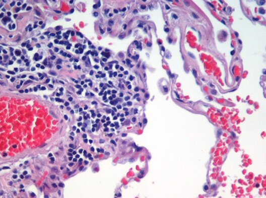
To counter this, the liver produces alpha-1 antitrypsin (AAT), an antiprotease. AAT circulates in the blood, travels to the lungs, and acts as a shield, specifically inhibiting neutrophil elastase. In AATD, particularly with the severe Z allele, the liver fails to secrete enough functional AAT. This creates a 'loss-of-function' scenario in the lungs. Without its inhibitor, neutrophil elastase runs rampant, systematically destroying the alveolar walls. This unchecked destruction leads to panacinar emphysema, where the air sacs lose their structure, enlarge, and merge, severely impairing gas exchange.
The Liver: A Toxic Buildup
While the lungs suffer from the absence of AAT, the liver suffers from its presence. The Z variant of the AAT protein has a mutation that causes it to misfold as it's being synthesized inside the liver cells (hepatocytes). Instead of being properly processed and secreted, these sticky, misshapen proteins get stuck inside the cell's protein-folding factory, the endoplasmic reticulum (ER).
Trapped in the ER, the Z-AAT proteins begin to link together, forming long chains or polymers. This is a 'toxic gain-of-function' mechanism. The accumulation of these polymers causes immense on the hepatocyte. The cell's machinery becomes overwhelmed trying to clear out the toxic buildup, leading to inflammation, cell death, and scarring (fibrosis). Over time, this chronic injury can progress to cirrhosis and even increase the risk for hepatocellular carcinoma.
Under a microscope, these intracellular polymers are visible as distinctive within the hepatocyte cytoplasm. Their presence is a hallmark sign of AATD-related liver disease.
A Disease of Two Timelines
Curiously, the liver disease caused by AATD often follows a bimodal distribution. A certain percentage of individuals present with liver problems, such as jaundice or elevated liver enzymes, in infancy and early childhood. Many of these cases resolve, but some can progress rapidly to liver failure.
For those who don't show early signs, the risk resurfaces in adulthood, typically after age 50. The reasons for this pattern aren't fully understood but may involve a 'second hit' hypothesis. The underlying proteotoxic stress from AAT polymerization may make the liver more vulnerable to other insults over a lifetime, such as viral infections, alcohol use, or metabolic issues, which can then trigger significant disease progression.
Let's test your understanding of how AATD affects both the lungs and the liver.
How does the genetic defect in Alpha-1 Antitrypsin Deficiency (AATD) cause damage to the lungs and liver, respectively?
In the context of AATD, the destruction of the alveolar walls leading to emphysema is a direct result of:
The dual pathophysiology of AATD, driven by loss-of-function in the lungs and toxic gain-of-function in the liver, highlights how one genetic error can lead to distinct and damaging outcomes in different parts of the body.