Clinical Management and Biomechanics of Foot Drop
Neural Pathways and Localization
From Spine to Foot
To understand foot drop, we need to trace the nerve signal from its origin. The journey begins at the L5 nerve root in your lower back. Think of this as a major highway exit. Fibers from L5, along with others from L4, S1, S2, and S3, merge to form the body's largest nerve: the sciatic nerve.
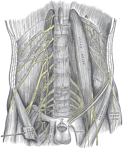
The sciatic nerve travels down the back of the thigh. It’s a bundle of two distinct nerves wrapped in a common sheath: the tibial and common peroneal nerves. Just above the back of the knee, in a space called the popliteal fossa, this bundle officially splits. The tibial nerve continues straight down, while our nerve of interest, the common peroneal nerve (also called the common fibular nerve), takes a detour.
A Vulnerable Pathway
The common peroneal nerve (CPN) wraps around the outside of the knee, right at the neck of the fibula—the thinner of the two lower leg bones. This path around the is a critical point. The nerve is very close to the surface here, with little muscle or fat to protect it from compression or direct injury. It's the most common site for nerve entrapment in the lower leg.
After rounding the fibular neck, the CPN divides into its two main terminal branches: the superficial peroneal nerve and the deep peroneal nerve.
Division of Labor
These two branches have very different jobs. The deep peroneal nerve dives deep into the anterior (front) compartment of the leg. It controls the muscles responsible for dorsiflexion (lifting the foot up) and toe extension. It also provides sensation to the small web of skin between the big toe and the second toe. Damage here is the classic cause of —the inability to lift the front part of the foot.
The superficial peroneal nerve stays more to the side, in the lateral compartment. It controls the muscles that evert the foot (turn the sole outwards). Its sensory role is much larger than its deep counterpart, providing feeling to the top of the foot and the front of the lower leg.
| Nerve Branch | Motor Function (Action) | Sensory Function (Feeling) |
|---|---|---|
| Deep Peroneal | Dorsiflexion (foot up) & Toe Extension | Web space between 1st & 2nd toes |
| Superficial Peroneal | Eversion (foot out) | Top of the foot (dorsum) |
A handy clinical mnemonic helps keep this straight: PED and TIP.
- PED: Peroneal nerve controls Eversion and Dorsiflexion.
- TIP: Tibial nerve controls Inversion and Plantarflexion.
By testing these specific movements, a clinician can quickly localize the problem to a specific nerve pathway.
Sometimes, a patient's symptoms can't be explained by a single point of injury. A nerve can be compressed at more than one location along its path, such as at the L5 root in the spine and at the fibular neck. This is known as the , where compression at one site makes the nerve more vulnerable to damage at another.
Now, let's test your knowledge on these neural pathways.
Where does the sciatic nerve typically divide into the tibial and common peroneal nerves?
A patient is unable to lift their foot upwards (dorsiflexion) and has numbness in the webbing between their big toe and second toe. Which nerve is most likely damaged?