Clinical Laboratory Result Interpretation
Acid Base Synthesis
Unmasking Complex Acid-Base Disorders
In critically ill patients, particularly those with multi-system organ failure, standard acid-base parameters can be dangerously misleading. A normal pH doesn't always signify homeostasis. It can, in fact, conceal a life-threatening
tug-of-war
between opposing acid-base disturbances. For instance, a patient with diabetic ketoacidosis (metabolic acidosis) who is also vomiting (metabolic alkalosis) might present with a deceptively normal pH and bicarbonate. Similarly, a patient with sepsis-induced lactic acidosis might hyperventilate, inducing a respiratory alkalosis that normalizes their pH. Relying solely on pH, pCO2, and HCO3− is insufficient in these scenarios. A deeper analysis is required to quantify each contributing process.
The Delta-Delta Calculation
When an elevated anion gap metabolic acidosis (HAGMA) is present, the next step is to determine if other metabolic disorders are lurking beneath the surface. The 'Delta-Delta' relationship, also known as the , is a crucial tool for this. It compares the change in the anion gap (ΔAG) to the change in bicarbonate (ΔHCO3−). In a pure HAGMA, for every 1 mEq/L increase in the anion gap, there should be a corresponding 1 mEq/L decrease in bicarbonate as it buffers the excess acid. The ratio quantifies this relationship.
Consider a patient with a sodium of 140, chloride of 100, and bicarbonate of 15. The anion gap is 25 mEq/L (140 - (100 + 15)), which is elevated. The ΔAG is 13 (25 - 12), and the ΔHCO3− is 9 (24 - 15). The Delta Ratio is 13/9 ≈ 1.44, consistent with a pure HAGMA. However, if the bicarbonate were 20, the ΔHCO3− would be 4, and the ratio would be 13/4 = 3.25. This points to a hidden metabolic alkalosis.
Adjusting for Albumin
The anion gap calculation itself has a significant weakness in the ICU: it's dependent on albumin, the main unmeasured anion in the blood. Hypoalbuminemia is rampant in critical illness due to malnutrition, inflammation, and fluid shifts. Since albumin is a negatively charged protein, a low level will artificially lower the calculated anion gap, potentially masking a serious HAGMA. To correct for this, we must calculate the albumin-adjusted anion gap.
Stewart vs. Henderson-Hasselbalch
The traditional Henderson-Hasselbalch approach treats bicarbonate as an independent variable, but in reality, it's a dependent variable influenced by other factors. The , developed by Peter Stewart, provides a more robust and quantitative model by focusing on three independent variables that truly determine the body's acid-base status.
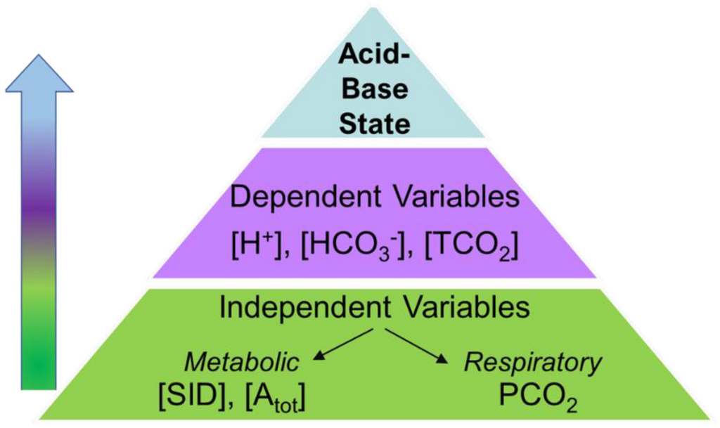
These independent variables are:
- The Strong Ion Difference (SID): The difference between all strong cations (Na+, K+, Ca2+, Mg2+) and strong anions (Cl−, lactate−) in plasma. A decrease in SID is acidifying, while an increase is alkalinizing.
- Total Weak Non-volatile Acids ([A_tot]): Primarily albumin and phosphate. An increase in [A_tot] (e.g., hyperphosphatemia in renal failure) is acidifying.
- Partial Pressure of Carbon Dioxide (PCO2): The respiratory component, same as in the traditional model.
This framework excels in quantifying the individual contributions to an acid-base disturbance. For instance, in a septic patient, the Stewart approach can separately measure the acidifying effect of lactate (decreasing SID), the alkalinizing effect of hypoalbuminemia (decreasing [A_tot]), and the compensatory respiratory alkalosis (decreasing PCO2). This is far more precise than simply diagnosing a 'mixed disorder'.
In patients with acute-on-chronic renal failure, these derangements become even more complex. Chronic acidosis leads to significant buffering by bone, and chronic hyperkalemia may be tolerated due to cellular adaptations. When an acute insult occurs, the resulting electrolyte shifts can be unpredictable, making the quantitative Stewart approach even more valuable for guiding therapy, such as the formulation of dialysis fluid.
A patient in the ICU has a normal pH of 7.40, a pCO2 of 25 mmHg, and a bicarbonate of 15 mEq/L. Based on these values alone, what is the most likely scenario?
Why is it crucial to calculate an albumin-adjusted anion gap in many critically ill patients?
By moving beyond basic parameters and applying these advanced analytical frameworks, clinicians can uncover and precisely quantify the multiple, often opposing, forces at play in the acid-base balance of a critically ill patient.