Clinical Anatomy of Trochanteric Anastomosis
Vascular Network Origins
The Arterial Inflow
The blood supply to the hip, specifically the head and neck of the femur, relies on a rich, interconnected network of arteries called an anastomosis. This network ensures that if one pathway is compromised, others can compensate. To understand this system, we first need to trace the main supply lines back to their origins.
Two major arteries descend from the abdominal aorta into the pelvis: the left and right common iliac arteries. Each one quickly divides, creating two distinct pathways relevant to the hip: the internal iliac artery, which supplies pelvic structures, and the external iliac artery, which continues down to supply the lower limb.
The Circumflex Arteries
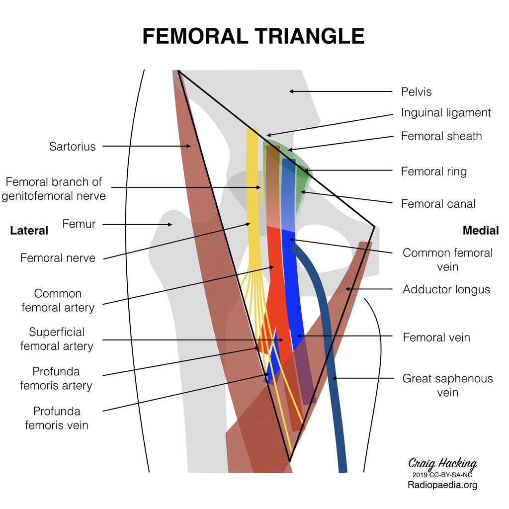
The primary blood supply to the femoral head comes from two key arteries that wrap around the femoral neck. These are the medial and lateral circumflex femoral arteries (MCFA and LCFA). Both typically arise from the deep femoral artery (also known as the profunda femoris), which is the largest branch of the femoral artery.
The Medial Circumflex Femoral Artery (MCFA) is arguably the most important vessel for the femoral head. After branching from the deep femoral artery, it travels posteriorly, passing between the iliopsoas and pectineus muscles, to wrap around the back of the femoral neck. Its branches pierce the joint capsule to supply the majority of the head and neck.
The Lateral Circumflex Femoral Artery (LCFA) travels in the opposite direction, running anteriorly and laterally. It passes deep to the sartorius and rectus femoris muscles, where it splits into ascending, transverse, and descending branches. The ascending branch is the one that contributes to the trochanteric anastomosis.
The Gluteal Arteries
The other major contributors to the hip's vascular network come from the internal iliac artery. These are the superior and inferior gluteal arteries, which exit the pelvis through the greater sciatic foramen to supply the muscles of the gluteal region.
Think of the circumflex arteries as the local specialists for the femoral head and neck, while the gluteal arteries are regional suppliers that also send crucial branches to the area.
The Superior Gluteal Artery (SGA) is the largest branch of the internal iliac artery. It exits the pelvis superior to the piriformis muscle. Its deep branches supply the gluteus medius and minimus, and it sends contributing vessels to the trochanteric anastomosis.
The Inferior Gluteal Artery (IGA) exits the pelvis inferior to the piriformis muscle, running alongside the sciatic nerve. It provides the main blood supply to the gluteus maximus muscle and also contributes a branch that ascends to join the network around the greater trochanter.
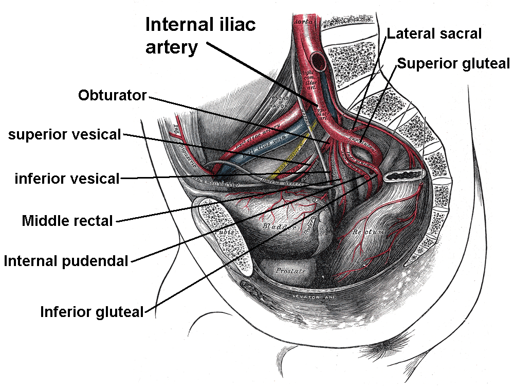
Together, these four arteries—MCFA, LCFA, SGA, and IGA—form the foundational supply for the trochanteric anastomosis. Their distinct origins from both the external and internal iliac systems create a redundant and resilient blood supply to one of the most critical joints in the body.
Which artery is considered the most important for supplying blood to the head and neck of the femur?
A disruption of blood flow in the deep femoral artery (profunda femoris) would most directly impact which two key arteries supplying the femoral neck?
Now that we've identified the suppliers, we can examine how they connect.