Clinical Anatomy of the Popliteal Fossa
Anatomical Boundaries
The Popliteal Fossa
Behind your knee is a shallow, diamond-shaped depression known as the popliteal fossa. It’s more than just a space; it's a crucial anatomical junction. Through this area pass major nerves, arteries, and veins that supply the lower leg and foot. Understanding its boundaries is key to diagnosing issues like cysts, aneurysms, and nerve entrapment.
Defining the Diamond
The popliteal fossa is framed by powerful muscles of the thigh and leg. These muscular borders give the space its distinct shape.
| Border | Muscle(s) |
|---|---|
| Superolateral (upper, outer) | Biceps femoris |
| Superomedial (upper, inner) | Semimembranosus and Semitendinosus |
| Inferolateral (lower, outer) | Lateral head of the gastrocnemius muscle |
| Inferomedial (lower, inner) | Medial head of the gastrocnemius muscle |
The two superior borders are formed by the distal ends of the hamstring muscles. The inferior borders are created by the two heads of the gastrocnemius, the primary muscle of the calf.
Floor and Roof
The fossa isn't an empty void; it has a distinct floor and roof that enclose the neurovascular structures running through it.
The floor, or the deep surface, is what you would feel if you could press through the nerves and vessels. It's composed of several structures from top to bottom:
- The popliteal surface of the femur
- The posterior capsule of the knee joint, reinforced by the oblique popliteal ligament
- The strong fascia covering the popliteus muscle
The roof is much simpler. It’s the superficial layer covering the diamond. It consists of the skin and a tough sheet of connective tissue called the popliteal fascia, which is a continuation of the thigh's deep fascia, the fascia lata.
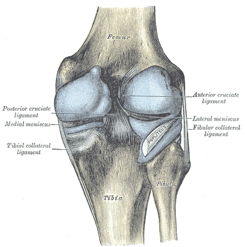
These boundaries are not just anatomical trivia. A common clinical issue, a Baker's cyst, is a fluid-filled sac that forms from the knee joint. Because the joint capsule is part of the fossa's floor, this cyst can bulge posteriorly, pushing through the roof between the gastrocnemius heads. The rigid boundaries of the fossa mean that any swelling or mass can compress the vital nerves and vessels within, leading to serious complications.
Let's check your understanding of these boundaries.
What forms the two inferior borders of the diamond-shaped popliteal fossa?
Which of the following structures is part of the 'floor' of the popliteal fossa?
Knowing the layout of the popliteal fossa provides a clear map for locating structures and understanding pathologies behind the knee.