Clinical Anatomy of the Popliteal Fossa
Popliteal Fossa Boundaries
The Popliteal Fossa
Posterior to the knee joint lies a crucial anatomical space known as the popliteal fossa. Shaped like a diamond, this shallow depression is more than just the back of the knee; it's a major conduit for the neurovascular structures passing between the thigh and the leg. Understanding its precise boundaries is fundamental for clinical examinations and for locating these vital components.
Defining the Diamond
The fossa's diamond shape is formed by the arrangement of several key muscles of the posterior thigh and leg. These muscular borders define the space and protect the contents within.
Think of the fossa as having two triangles: an upper one formed by the hamstring muscles and a lower one formed by the calf muscle heads.
The superior, or upper, boundaries are created by the distal ends of the hamstring muscles.
- Superolateral margin: The biceps femoris muscle forms the upper-outer wall.
- Superomedial margin: The semimembranosus and, more superficially, the semitendinosus muscles define the upper-inner wall.
The inferior, or lower, boundaries are formed by the two heads of the most superficial calf muscle.
- Inferolateral margin: The lateral head of the gastrocnemius muscle.
- Inferomedial margin: The medial head of the gastrocnemius muscle. The small plantaris muscle also contributes to this border, lying deep to the gastrocnemius.
Floor and Roof
Beyond its muscular walls, the popliteal fossa has a deep floor and a superficial roof.
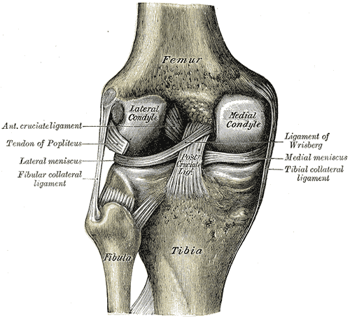
The floor, forming the base of the diamond, is not flat. From superior to inferior, it is composed of:
- The popliteal surface of the femur.
- The posterior capsule of the knee joint.
- The strong fascia covering the popliteus muscle.
The roof is much simpler. It consists of the skin and the dense deep fascia of the leg, known as the popliteal fascia. This fascia is pierced by a key vessel: the small saphenous vein. This vein runs up the back of the leg and passes through the fascial roof to drain into the popliteal vein, which lies within the fossa.
| Boundary | Structures |
|---|---|
| Superolateral | Biceps femoris muscle |
| Superomedial | Semimembranosus & Semitendinosus muscles |
| Inferolateral | Lateral head of Gastrocnemius muscle |
| Inferomedial | Medial head of Gastrocnemius muscle |
| Floor (deep) | Popliteal surface of femur, knee joint capsule, popliteus fascia |
| Roof (superficial) | Skin & Popliteal fascia |
Let's check your understanding of these boundaries.
Which muscle forms the superolateral boundary of the popliteal fossa?
The inferior boundaries of the diamond-shaped popliteal fossa are formed by the two heads of which muscle?
Recognising these boundaries is the first step in navigating the complex anatomy of the posterior knee.