Clinical Anatomy of the Peroneal Trochlea
Osseous Morphology
The Peroneal Trochlea
On the lateral side of the calcaneus, or heel bone, sits a small but significant bony prominence known as the peroneal trochlea (PT). Also called the peroneal tubercle, this structure is a key landmark for understanding the function and potential pathologies of the lateral ankle and foot. It's typically found in the middle third of the calcaneus's lateral surface.
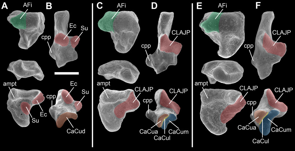
The primary role of the peroneal trochlea is to act as a separator. It functions like a pulley, creating two distinct pathways for the tendons of the fibularis muscles. The tendon of the muscle passes through a groove superior to the trochlea, while the tendon of the fibularis longus muscle runs in a groove inferior to it. This bony partition prevents the tendons from rubbing against each other and ensures they maintain an efficient line of pull during movement.
This entire complex is stabilised by the inferior peroneal retinaculum, a band of connective tissue. A septum from this retinaculum actually inserts directly onto the peroneal trochlea, reinforcing the separation of the two tendon compartments. The trochlea's location is also consistently anterior to the attachment point of the calcaneofibular ligament, one of the main lateral stabilisers of the ankle.
Posterior to the peroneal trochlea is another, usually less prominent, bony elevation called the (RTE). The space between the PT and the RTE forms the channel through which the tendons pass. The relative size and relationship between these two structures can influence tendon stability.
Morphological Variations
The shape and size of the peroneal trochlea vary considerably among individuals. Researchers have categorised its morphology into four main types based on appearance. The conical and straight ridge types are by far the most common, each appearing in about 44% of the population.
| Type | Description | Incidence |
|---|---|---|
| Flat | The trochlea is barely a visible prominence. | ~7.2% |
| Conical | A distinct, cone-shaped projection. | ~44.0% |
| Straight Ridge | A linear, ridge-like elevation. | ~44.6% |
| Olecranon-type | A large, prominent, hook-like process. | ~4.2% |
While most variations are asymptomatic, a particularly large or misshapen peroneal trochlea, such as a prominent olecranon-type, can lead to clinical problems. This condition, known as peroneal trochlear hypertrophy, can cause chronic pain, irritation (tendinopathy), or even tearing of the fibularis longus tendon as it is forced to take a sharp turn around the enlarged bone. In such cases, surgical reduction of the trochlea may be required to relieve the impingement on the tendon.
What is the primary function of the peroneal trochlea?
The tendon of the fibularis brevis muscle passes _______ the peroneal trochlea, while the tendon of the fibularis longus passes _______.
Understanding the anatomy of the peroneal trochlea and its variations is crucial for diagnosing and managing lateral ankle pain and instability.