Clinical Anatomy and Surgical Significance of the Trochanteric Anastomosis
Anastomosis Structural Components
The Trochanteric Anastomosis
At the back of the hip joint, nestled in a small depression on the femur called the trochanteric fossa, lies a critical network of blood vessels. This network, the trochanteric anastomosis, is a meeting point for several arteries. Its primary job is to ensure a steady blood supply to the head of the femur, the rounded top part that fits into the hip socket.
Think of it as a roundabout for blood flow. If one road is blocked, traffic can still get through via the other connecting roads. This redundancy is vital for maintaining the health of the femoral head bone tissue. Without it, the bone could be at risk of avascular necrosis, a condition where the bone tissue dies due to a lack of blood.
This network is formed by four main contributors.
The Four Contributors
The trochanteric anastomosis isn't a single vessel, but a junction of branches from four separate arteries.
-
Ascending branch of the Medial Circumflex Femoral Artery (MCFA): This is the most important player. The MCFA provides the majority of the blood to the femoral head. Its ascending branch travels up the back of the femoral neck to join the anastomosis.
-
Ascending branch of the Lateral Circumflex Femoral Artery (LCFA): This artery approaches from the other side. Its ascending branch contributes to the network, providing an alternative route for blood flow.
-
Superior Gluteal Artery: This artery primarily supplies the gluteal muscles, but it sends a branch down to connect with the other vessels in the trochanteric fossa.
-
Inferior Gluteal Artery: Similar to its superior counterpart, this artery also supplies the gluteal region and contributes a branch to the anastomosis, completing the network.
The majority of the blood supply to the femoral head is derived from the extracapsular arterial ring formed by the medial and lateral femoral circumflex arteries, which arise from the deep femoral artery.
The Medial and Lateral Circumflex Femoral Arteries form an arterial ring around the base of the femoral neck. From this ring, smaller arteries, called retinacular arteries, ascend along the femoral neck within the joint capsule to penetrate and nourish the femoral head. The gluteal arteries provide additional, secondary connections to this system.
Topography of the Fossa
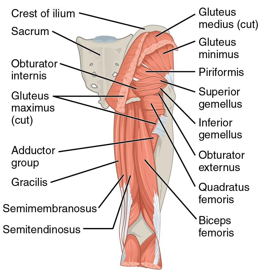
The precise location of this network is what makes it so effective. The trochanteric fossa is a deep depression on the medial surface of the greater trochanter. This bony landmark provides a protected space for these crucial arteries to connect.
The vessels enter this space and are situated close to the hip joint capsule. This proximity allows for the formation of the vascular ring that ultimately feeds the femoral head. The anastomosis acts as a critical backup system, ensuring that if flow from one direction is reduced, blood can still reach the femoral head from the other contributing arteries, preserving the joint's integrity.
Time to check your understanding of this crucial vascular network.
What is the primary function of the trochanteric anastomosis?
Which of the following arteries is NOT one of the four main contributors to the trochanteric anastomosis?
Understanding this anatomy is crucial for grasping how the hip joint is sustained and how it can be vulnerable to injury.