Clinical Anatomy and Functional Dynamics of the Gluteal Region
Gluteal Spatial Organization
The Gluteal Architecture
The gluteal region is more than just a group of muscles; it's a complex, layered structure critical for movement and stability. Its architecture begins with the skin and underlying fascial layers. The superficial fascia contains a significant amount of adipose tissue, forming the gluteal fat pads. The volume and distribution of this fat vary, but it provides cushioning and contour to the region.
Beneath this is the deep fascia, or fascia lata, a strong fibrous sheet that encases the thigh muscles. Superiorly, it splits to enclose the gluteus maximus and tensor fasciae latae, forming distinct compartments that are clinically important as they can contain the spread of fluid or infection.
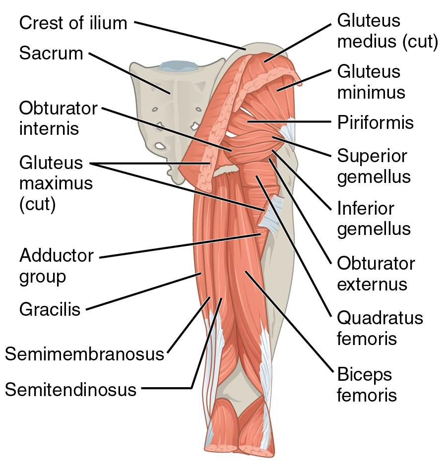
The muscles themselves are organised into two main layers. The most superficial layer is dominated by the large gluteus maximus. It covers most of the other gluteal muscles. When the gluteus maximus is reflected, the deeper layer is revealed, primarily consisting of the gluteus medius and, deep to that, the gluteus minimus. This layered arrangement is key to their coordinated function in hip movement and pelvic stabilisation.
Beneath it lie the gluteus medius and minimus, which are crucial for thigh abduction and the stabilization of the pelvis during walking.
Functional Slings and Spaces
The gluteal muscles don't work in isolation. They form part of a functional system called the 'posterior oblique sling'. This myofascial connection links the gluteus maximus on one side with the latissimus dorsi on the opposite side via the thoracolumbar fascia. When you walk or run, this sling helps transfer force through the pelvis, stabilising the sacroiliac joint and providing power for propulsion.
The iliotibial (IT) band is another critical structure. It's not a muscle, but a thickened lateral portion of the fascia lata. The gluteus maximus and the tensor fasciae latae (TFL) both insert into the IT band, placing it under tension. This tension is vital for stabilising the knee and hip during the stance phase of gait, preventing the pelvis from dropping on the unsupported side.
Between these muscle and fascial layers are potential spaces known as bursae. A bursa is a fluid-filled sac that reduces friction between tissues. In the gluteal region, three are particularly important:
- Trochanteric Bursa: Located between the greater trochanter of the femur and the gluteus maximus. Inflammation here causes pain on the side of the hip.
- Ischial Bursa: Lies between the ischial tuberosity and the gluteus maximus. It can become inflamed from prolonged sitting.
- Gluteofemoral Bursa: Separates the IT band from the vastus lateralis muscle.
Understanding the location of these bursae is essential, as they are common sites of pathology. Inflammation, or bursitis, can mimic other conditions like sciatica or hip joint arthritis.
Defining the Boundaries
To accurately locate deeper structures, we must first define the spatial boundaries of the gluteal region.
- Superiorly: The iliac crest forms the upper boundary.
- Medially: The intergluteal cleft (the groove between the buttocks) is the medial border.
- Inferiorly: The gluteal fold, the horizontal crease at the bottom of the buttock, marks the lower limit.
- Laterally: The region extends to a line drawn from the anterior superior iliac spine (ASIS) to the greater trochanter.
These surface landmarks correspond to the underlying musculoskeletal anatomy. The volume within these borders contains not only the gluteal muscles, fat, and bursae, but also the critical neurovascular bundles that pass from the pelvis into the lower limb. A firm grasp of this three-dimensional organisation is the foundation for safe clinical procedures and accurate diagnosis in this complex region.
Which of the following correctly lists the layers of the gluteal region from superficial to deep?
The posterior oblique sling describes a functional myofascial connection between which two muscles?