CCRN Mastery and Critical Care Excellence
Advanced Hemodynamic Integration
Pulmonary Artery Catheter Waveforms
The pulmonary artery (PA) catheter, or Swan-Ganz, gives us a detailed look inside the heart and pulmonary circulation. Beyond simple pressures, the waveform itself tells a story about right heart function. When looking at a pulmonary artery occlusion pressure (PAOP) or wedge pressure waveform, you'll see three distinct waves corresponding to events in the right atrium.
Let's break down what each wave means:
- 'a' wave: This reflects atrial contraction, or 'atrial kick.' It's the rise in pressure as the right atrium squeezes blood into the right ventricle. It lines up with the PR interval on an EKG.
- 'c' wave: A smaller wave caused by the bulging of the tricuspid valve back into the right atrium during early ventricular contraction (isovolumetric contraction). It occurs after the QRS complex.
- 'v' wave: This represents atrial filling. As the atrium passively fills with blood from the body while the tricuspid valve is closed, pressure rises, creating the 'v' wave. It ends just as the tricuspid valve opens.
An absent 'a' wave can indicate atrial fibrillation, since there is no coordinated atrial contraction. Giant 'v' waves might suggest tricuspid regurgitation, as blood flows backward into the atrium during ventricular systole.
Troubleshooting the Catheter
PA catheter readings are only useful if they're accurate. Two common problems are dampened waveforms and accidental wedging.
A dampened waveform looks flattened, with a loss of the clear dicrotic notch and distinct systolic and diastolic pressures. This can be caused by an air bubble in the tubing, a loose connection, a clot at the catheter tip, or the catheter tip pressing against a vessel wall. The first steps are to check your setup: ensure all connections are tight, flush the line to dislodge potential clots or bubbles, and check that the pressure bag is inflated correctly. If these steps don't work, the patient may need to change position.
An accidental wedge occurs when the catheter migrates further into a smaller pulmonary artery branch, occluding it. The waveform will change from a distinct PA systolic/diastolic pattern to a lower-pressure PAOP waveform. This is a medical emergency because it can cause a pulmonary infarct if left unresolved. The nurse's immediate action is to attempt to reposition the patient. If that fails, a physician or advanced practice provider should be notified immediately to pull the catheter back slightly.
Measuring Cardiac Output and Resistance
The PA catheter allows us to measure cardiac output (CO) using the thermodilution method. A small amount of room-temperature or iced saline is injected rapidly into the proximal (right atrial) port. A thermistor at the catheter's distal tip measures the change in blood temperature as the cooler saline mixes and flows past. The machine's computer analyzes the temperature change over time, displayed as a thermodilution curve, and calculates the cardiac output.
A smooth, rapid curve indicates a good injection and a healthy CO. A slow, drawn-out curve can suggest a low cardiac output, as the blood is moving sluggishly. Limitations include inaccurate readings in patients with tricuspid regurgitation (which causes injectate to flow backward) or intracardiac shunts.
From cardiac output and pressure measurements, we can calculate vascular resistance. This tells us how much opposition the ventricle must overcome to push blood forward. There are two key types.
Afterload
noun
The pressure or resistance the heart must overcome to eject blood during systole. It's the 'load' the heart must work against.
Systemic Vascular Resistance (SVR) reflects the afterload of the left ventricle. It's the total resistance of the entire body's arterial system. Pulmonary Vascular Resistance (PVR) is the afterload of the right ventricle, representing the resistance within the pulmonary circulation.
These are not measured directly but are calculated using hemodynamic values.
A high SVR means the left ventricle is working hard to pump blood to the body, often seen with vasoconstriction. A high PVR indicates the right ventricle is struggling to pump blood into the lungs, a hallmark of pulmonary hypertension.
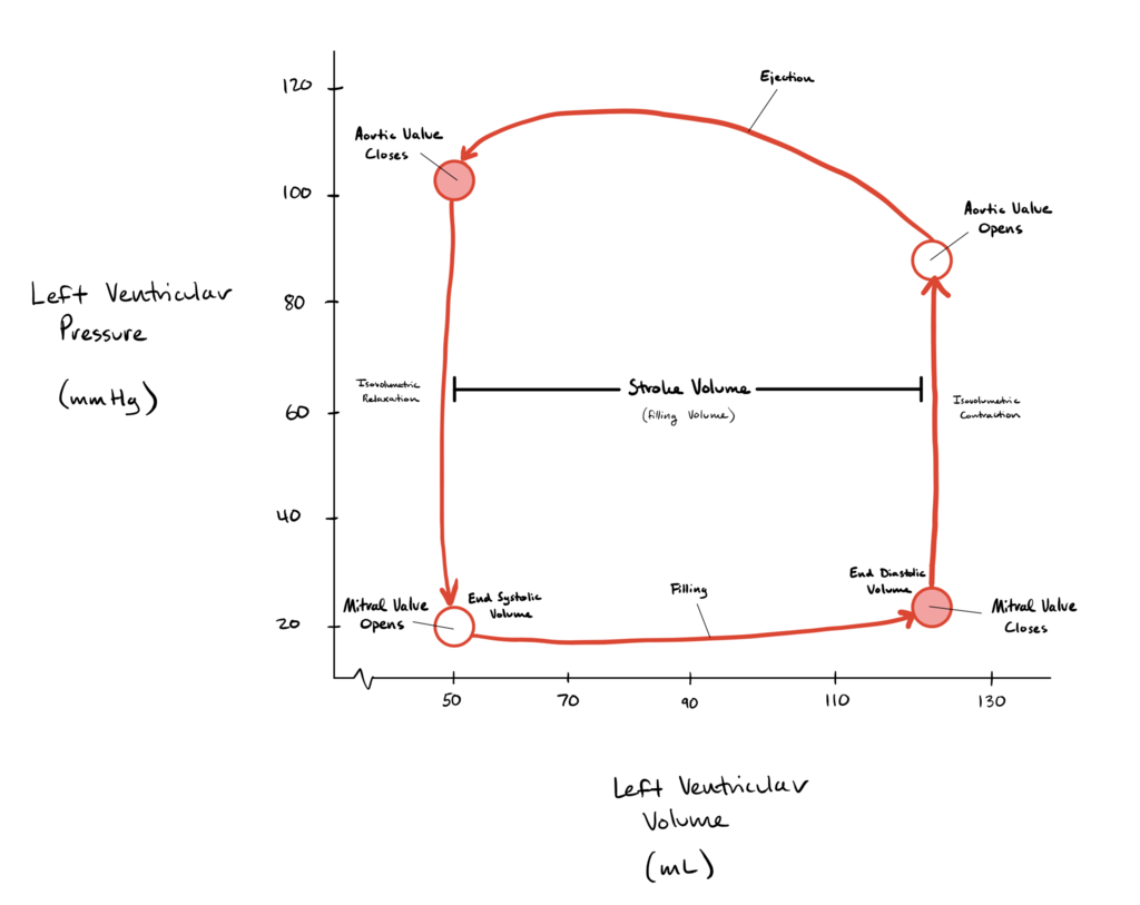
LV stroke volume creates arterial pulse pressure by distending conducting vessels during systole, and SVR preserves diastolic pressure by preventing SV from flowing through arterioles during diastole. This Ventricular-vascular coupling allows rapid clinical separation of hypotensive patients into those with increased SV and CO ... and those with ... low SV, low CO, increased SVR (cardiogenic, hypovolaemic shock).
Putting It All Together
By integrating these values, we can build a hemodynamic profile to identify different types of shock and differentiate between right and left heart failure.
For example, the relationship between Pulmonary Artery Diastolic (PAD) pressure and PAOP is key. In a healthy heart, PAD is slightly higher than PAOP ( mmHg). If the gradient is large (e.g., PAD is mmHg and PAOP is mmHg), it suggests a problem in the lungs, like pulmonary hypertension or a pulmonary embolism, which increases PVR. This points to right heart failure.
Conversely, if both PAD and PAOP are high and roughly equal, the problem is likely originating from the left side of the heart. The left ventricle is failing to pump effectively, causing blood to back up into the pulmonary circulation and increase pressures throughout. This is indicative of left heart failure.
Different shock states also have distinct hemodynamic profiles. Understanding these patterns is critical for guiding therapy, such as choosing between vasopressors (to increase SVR) and inotropes (to improve contractility and CO).
| Shock Type | Preload (CVP/PAOP) | Afterload (SVR) | Cardiac Output/Index | Clinical Picture |
|---|---|---|---|---|
| Hypovolemic | Low | High | Low | Empty tank; body clamps down to compensate. |
| Cardiogenic | High | High | Low | Pump failure; blood backs up, body clamps down. |
| Distributive (Septic) | Low (initially) | Low | High (initially) | Leaky, dilated pipes; heart pumps fast but ineffectively. |
In a clinical scenario, if a patient is hypotensive with a very low SVR and a high CO, the diagnosis points toward distributive shock. The appropriate intervention is a vasopressor like norepinephrine to increase SVR and restore vessel tone. If another patient has high CVP and PAOP, high SVR, and a low CO, this profile fits cardiogenic shock, and treatment would focus on inotropes like dobutamine to improve contractility.
Ready to test your knowledge?
When analyzing a pulmonary artery occlusion pressure (PAOP) waveform, which wave corresponds to atrial filling while the tricuspid valve is closed?
A nurse notices a patient's pulmonary artery pressure waveform has become flattened, with loss of the dicrotic notch. What is the priority action?
Mastering hemodynamic interpretation is a core skill in critical care, allowing you to see beyond basic vital signs and tailor therapies to the underlying physiology.