Cardiology for Medical Students
Cardiovascular Anatomy
The Four-Chambered Pump
The heart is the engine of the circulatory system, a powerful muscle organized into four distinct chambers. Think of it as a duplex apartment, with two rooms on the right and two on the left, separated by a thick muscular wall called the septum. This separation is crucial because the right side of the heart handles deoxygenated blood, while the left side manages oxygenated blood. Mixing them would make circulation inefficient.
The two upper chambers are the atria (singular: atrium), which act as receiving rooms for blood entering the heart. The two lower chambers are the ventricles, the powerhouses that pump blood out to the lungs and the rest of the body.
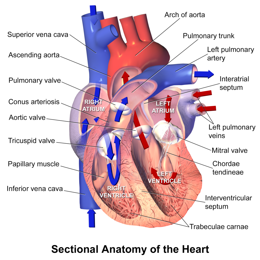
Here's the path blood takes:
- Right Atrium: Receives deoxygenated blood from the body through two large veins, the superior and inferior vena cava.
- Right Ventricle: Pumps this deoxygenated blood to the lungs.
- Left Atrium: Receives oxygen-rich blood from the lungs.
- Left Ventricle: Pumps this oxygenated blood out to the entire body through the aorta.
Notice the muscular wall of the left ventricle is much thicker than the right. That's because it needs to generate significantly more force to push blood to every part of your body, from your brain to your toes. The right ventricle only needs to pump blood next door to the lungs.
One-Way Gates
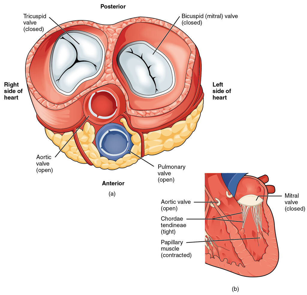
To keep blood flowing in the right direction, the heart has four valves. These valves are like one-way doors that open to let blood pass through and then snap shut to prevent it from flowing backward.
The valves work in pairs. Two valves, called the atrioventricular (AV) valves, control blood flow from the atria to the ventricles.
Tricuspid Valve: Located between the right atrium and right ventricle. It has three flaps, or cusps.
Mitral Valve (or Bicuspid Valve): Located between the left atrium and left ventricle. It has two cusps.
The other two valves, the semilunar valves, control blood flow out of the ventricles and into the major arteries. They are named for their half-moon-shaped cusps.
Pulmonary Valve: Located between the right ventricle and the pulmonary artery.
Aortic Valve: Located between the left ventricle and the aorta.
Feeding the Heart Itself
The heart muscle, or myocardium, is a relentless worker that needs its own dedicated supply of oxygen and nutrients. This is the job of the coronary circulation system, a network of arteries and veins that wrap around the outside of the heart.
The main pipelines are the coronary arteries, which branch off the aorta right after it leaves the left ventricle. This positioning ensures they receive the most oxygen-rich blood under the highest pressure.
The two primary coronary arteries are:
- Right Coronary Artery (RCA): Supplies blood mainly to the right side of the heart.
- Left Main Coronary Artery (LMCA): This is a short vessel that quickly divides into two major branches:
- Left Anterior Descending (LAD) Artery: Supplies the front and bottom of the left ventricle and the front of the septum.
- Circumflex Artery (CX): Wraps around to supply blood to the back and side of the heart.
After the heart muscle uses the oxygen, the deoxygenated blood is collected by a system of cardiac veins. These veins generally run alongside the arteries and eventually drain into the coronary sinus, a large vein on the back of the heart that empties directly into the right atrium.
Let's check your understanding of these structures.
What is the primary function of the septum in the heart?
Which chamber of the heart has the thickest muscular wall, and why?
Understanding this basic layout of chambers, valves, and vessels is the first step to appreciating how this incredible organ works.