Cardiac Surgery Explained
Cardiac Anatomy
The Heart's Protective Sac
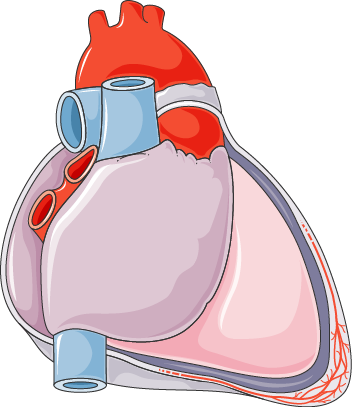
The heart sits inside a protective, fluid-filled sac called the pericardium. Think of it as a durable, double-layered bag. The outer layer, the fibrous pericardium, is tough and anchors the heart to surrounding structures, preventing it from overfilling with blood. The inner layer, the serous pericardium, is thinner and produces a lubricating fluid.
This pericardial fluid allows the heart to beat in a nearly frictionless environment, reducing wear and tear with every contraction.
pericardium
noun
The membrane enclosing the heart, consisting of an outer fibrous layer and an inner double layer of serous membrane.
Between the heart muscle itself and the pericardium lies the epicardium, which is actually the innermost layer of the pericardium. It contains coronary blood vessels and fat. Beneath that is the myocardium, the thick muscular wall of the heart responsible for pumping. The innermost layer is the endocardium, a thin lining that ensures blood flows smoothly through the heart's chambers.
A Four-Chambered Pump
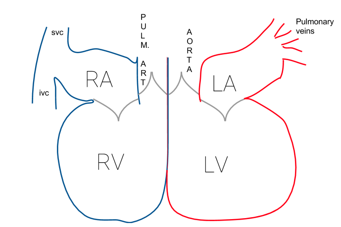
The heart is essentially two pumps working in unison. Each pump has two chambers: a receiving chamber called an atrium and a pumping chamber called a ventricle. The right side of the heart handles deoxygenated blood, while the left side manages oxygenated blood.
- Right Atrium: Receives deoxygenated blood from the body through two large veins, the superior and inferior vena cava.
- Right Ventricle: Pumps this deoxygenated blood to the lungs through the pulmonary artery.
- Left Atrium: Receives oxygen-rich blood from the lungs via the pulmonary veins.
- Left Ventricle: The strongest chamber. It pumps oxygenated blood to the rest of the body through the aorta. Its muscular wall is much thicker than the right ventricle's because it needs to generate more pressure to push blood throughout the entire body.
Valves and Vessels
To keep blood moving in the right direction, the heart has four one-way valves. They open to let blood pass through and snap shut to prevent it from flowing backward.
| Valve Name | Location | Function |
|---|---|---|
| Tricuspid Valve | Between the right atrium and right ventricle | Prevents backflow into the right atrium. |
| Pulmonary Valve | Between the right ventricle and pulmonary artery | Prevents backflow into the right ventricle. |
| Mitral Valve | Between the left atrium and left ventricle | Prevents backflow into the left atrium. |
| Aortic Valve | Between the left ventricle and the aorta | Prevents backflow into the left ventricle. |
The heart is also the hub for the body's largest blood vessels, often called the great vessels.
- Aorta: The largest artery in the body. It carries oxygenated blood from the left ventricle out to the systemic circulation.
- Pulmonary Artery: Carries deoxygenated blood from the right ventricle to the lungs.
- Vena Cavae: The superior and inferior vena cava are the two largest veins that bring deoxygenated blood from the body back to the right atrium.
- Pulmonary Veins: Unlike most veins, they carry oxygenated blood, returning it from the lungs to the left atrium.
Feeding the Heart Itself
The heart muscle is too thick to get its oxygen and nutrients from the blood passing through its chambers. It needs its own dedicated blood supply. This is the job of the coronary circulation system.
The coronary arteries branch off the aorta near the point where the aorta leaves the left ventricle. They wrap around the outside of the heart, delivering oxygen-rich blood to the myocardium. The main coronary arteries are the right coronary artery (RCA) and the left main coronary artery, which quickly splits into two major branches: the left anterior descending (LAD) and the circumflex artery.
Blockages in these crucial arteries are what cause heart attacks, as they deprive a section of the heart muscle of oxygen.
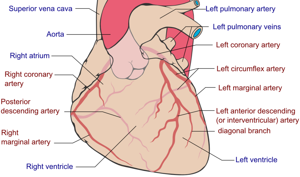
After the heart muscle has used the oxygen, deoxygenated blood is collected by cardiac veins. These veins run alongside the arteries and drain into the coronary sinus, a large vein on the back of the heart that empties directly into the right atrium.
Now, let's test your knowledge of the heart's anatomy.
What is the primary function of the pericardium?
Which chamber of the heart has the thickest muscular wall and why?
Understanding these structures—from the protective pericardium to the intricate network of coronary arteries—is the foundation for comprehending how the heart functions and how surgical interventions can address problems within this remarkable organ.