Cardiac Surgery Essentials
Cardiac Anatomy
The Heart’s Blueprint
The heart is a powerful, muscular pump at the center of the circulatory system. Think of it as a four-room house, with two rooms on the right and two on the left. The top rooms are called atria (singular: atrium), and the bottom rooms are called ventricles. A muscular wall called the septum divides the right side of the heart from the left, preventing blood from mixing.
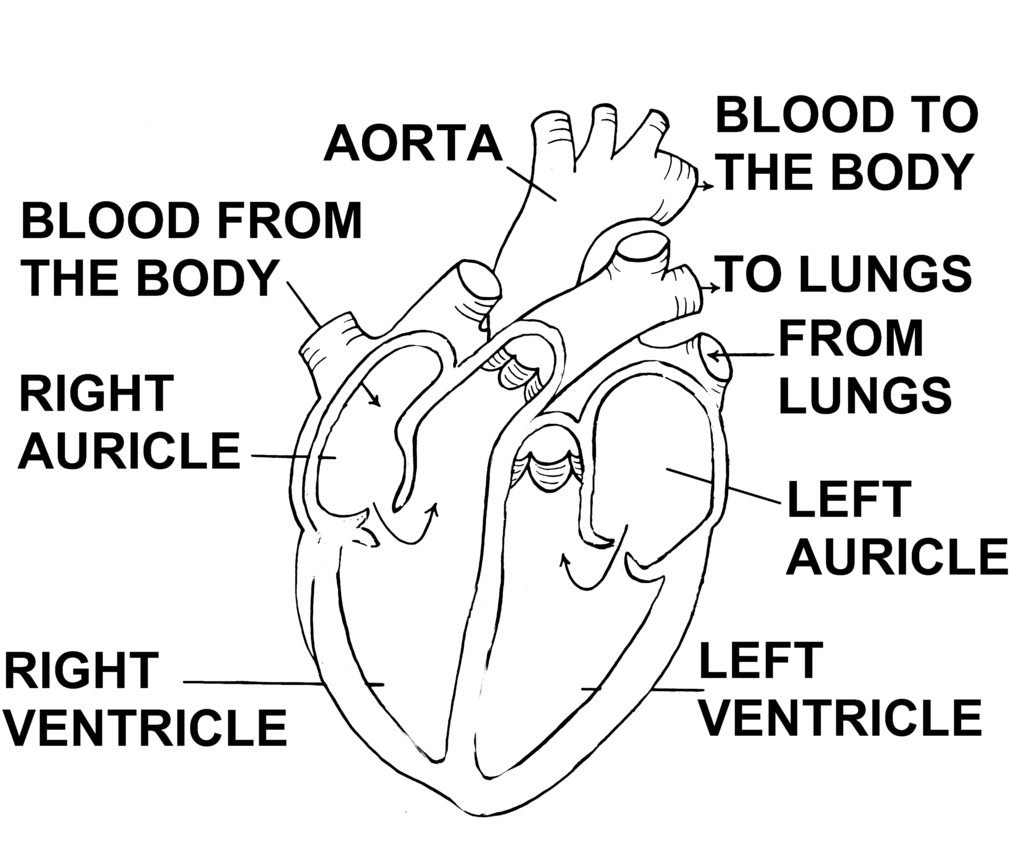
The right atrium receives deoxygenated blood—blood that has delivered oxygen to the body and is returning for a refill. From here, it passes into the right ventricle, which then pumps this blood to the lungs.
After picking up oxygen in the lungs, the blood flows into the left atrium. The left atrium sends this freshly oxygenated blood to the left ventricle. The left ventricle is the strongest chamber of the heart; its thick muscular wall contracts forcefully to pump oxygen-rich blood out to the rest of the body.
Right side: deoxygenated blood to the lungs. Left side: oxygenated blood to the body.
One-Way Doors
To keep blood flowing in the right direction, the heart has four valves that act like one-way doors. They open to let blood pass through and snap shut to prevent it from flowing backward.
There are two main types of valves:
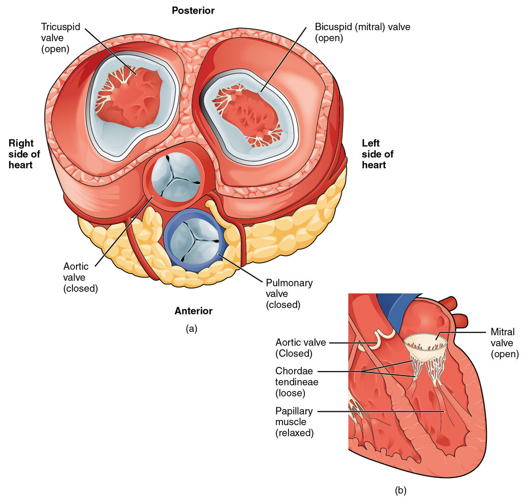
Atrioventricular (AV) Valves: These are located between the atria and ventricles.
- The tricuspid valve is between the right atrium and right ventricle.
- The mitral valve (or bicuspid valve) is between the left atrium and left ventricle.
Semilunar (SL) Valves: These are located at the exit of each ventricle, where blood leaves the heart.
- The pulmonary valve is at the exit of the right ventricle, opening into the pulmonary artery.
- The aortic valve is at the exit of the left ventricle, opening into the aorta.
Major Highways
Several large blood vessels are connected to the heart, acting as major highways for blood transport.
| Vessel | Connects to | Function |
|---|---|---|
| Superior & Inferior Vena Cava | Right Atrium | Bring deoxygenated blood from the body to the heart. |
| Pulmonary Artery | Right Ventricle | Carries deoxygenated blood from the heart to the lungs. |
| Pulmonary Veins | Left Atrium | Bring oxygen-rich blood from the lungs to the heart. |
| Aorta | Left Ventricle | The body's largest artery, it carries oxygen-rich blood from the heart to the rest of the body. |
This entire path, from the body to the heart, to the lungs, back to the heart, and out to the body again, is a continuous loop. The right side of the heart handles the pulmonary circuit (to the lungs), while the left side handles the systemic circuit (to the body).
Fueling the Pump
The heart muscle, or myocardium, works tirelessly and needs its own dedicated blood supply to get oxygen and nutrients. This is provided by the coronary circulation system. The coronary arteries branch off the aorta near the heart and wrap around the surface of the heart muscle.
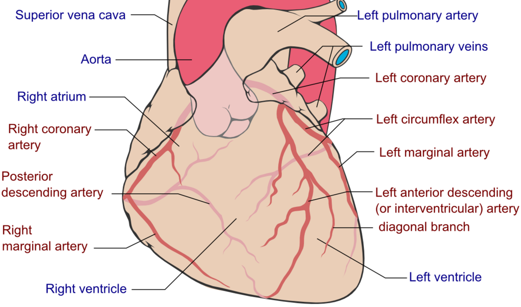
The two main coronary arteries are the left coronary artery and the right coronary artery. They divide into smaller branches that penetrate deep into the heart muscle, ensuring every cell receives the blood it needs to function. After the heart muscle uses the oxygen, coronary veins collect the deoxygenated blood and return it to the right atrium.
The Protective Sac
The heart is enclosed in a double-walled sac called the pericardium. The outer layer is a tough, fibrous sac that anchors the heart to surrounding structures and prevents it from overfilling with blood. The inner layer is a thin, delicate membrane.
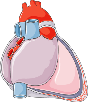
Between these two layers is a small amount of pericardial fluid. This fluid acts as a lubricant, allowing the heart to beat smoothly within the sac without creating friction. It’s a simple but elegant system that protects the heart as it works.
Now, let's test your knowledge of the heart's anatomy.
Deoxygenated blood from the body enters the heart through which chamber?
What is the primary function of the septum in the heart?
Understanding these fundamental components of cardiac anatomy is the first step in appreciating the complexity and elegance of the heart's function.