Cardiac Anatomy and the Dynamics of Blood Flow
Heart Chambers and Walls
The Heart's Four Rooms
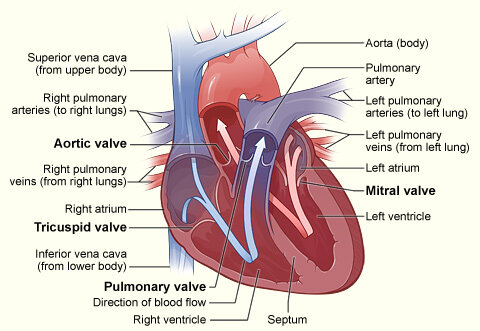
The heart isn't just a single pump; it's a duplex. It contains four distinct chambers that work in two coordinated pairs. The top two chambers are the atria (singular: atrium), which act as receiving rooms for blood returning to the heart. The bottom two are the ventricles, the powerful pumps that send blood out on its journey.
This four-chambered design allows the heart to manage two separate circulatory loops simultaneously: one to the lungs to pick up oxygen, and another to the rest of the body to deliver it. The right atrium and right ventricle handle deoxygenated blood, while the left atrium and left ventricle manage oxygenated blood. Keeping these two types of blood from mixing is critical for efficiency.
Built for the Job
The walls of these chambers aren't all created equal. Their structure is directly related to their function. The heart wall itself is primarily composed of a special type of muscle tissue called the . This is the engine of the heart, responsible for the powerful contractions that pump blood. Lining the inside of the chambers is a thin, smooth membrane called the endocardium, which ensures blood flows without friction.
The atria have relatively thin myocardial walls. Their job is simple: they just need to contract enough to push blood down into the ventricles directly below them. It's a short, low-pressure trip.
The ventricles, on the other hand, have much thicker, stronger walls. They are the heavy lifters. But even between the two ventricles, there's a significant difference. The right ventricle pumps deoxygenated blood to the lungs, a relatively short distance. The left ventricle has the most demanding job of all: it must pump oxygenated blood to every other part of the body, from your brain to your toes. This requires immense force.
Because it pumps blood to the entire body, the left ventricle's myocardium is the thickest and most muscular part of the heart—up to three times thicker than the right ventricle's wall.
The Dividing Walls
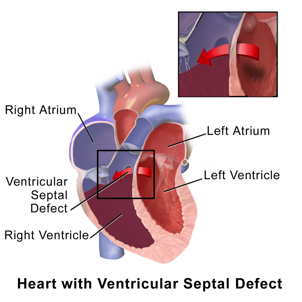
To keep the oxygen-rich and oxygen-poor blood completely separate, the heart is divided down the middle by a muscular wall known as the septum. The portion between the atria is the thin interatrial septum, while the much thicker wall between the ventricles is the .
This separation is fundamental to the efficiency of our circulatory system. It ensures that the blood going out to the body is fully oxygenated, maximizing the energy supplied to our cells. Any breach in this wall can cause serious problems by allowing the two types of blood to mix.
Ready to check your understanding of the heart's architecture?
Which chambers of the heart are primarily responsible for receiving blood returning from the body and lungs?
The right atrium and right ventricle are responsible for handling what type of blood?
The structure of each part of the heart is perfectly tailored to its role in the tireless process of circulating blood. The thin atria receive, the mighty ventricles pump, and the sturdy septum divides, all working together in a beautifully efficient system.