Canine Cataract Surgery Techniques
Canine Eye Anatomy
The Eye's Inner Workings
To understand cataracts, we first need to look at the basic anatomy of a dog's eye. While we'll focus on dogs, their eyes are remarkably similar to our own. Think of the eye as a biological camera, gathering light and turning it into images the brain can process.
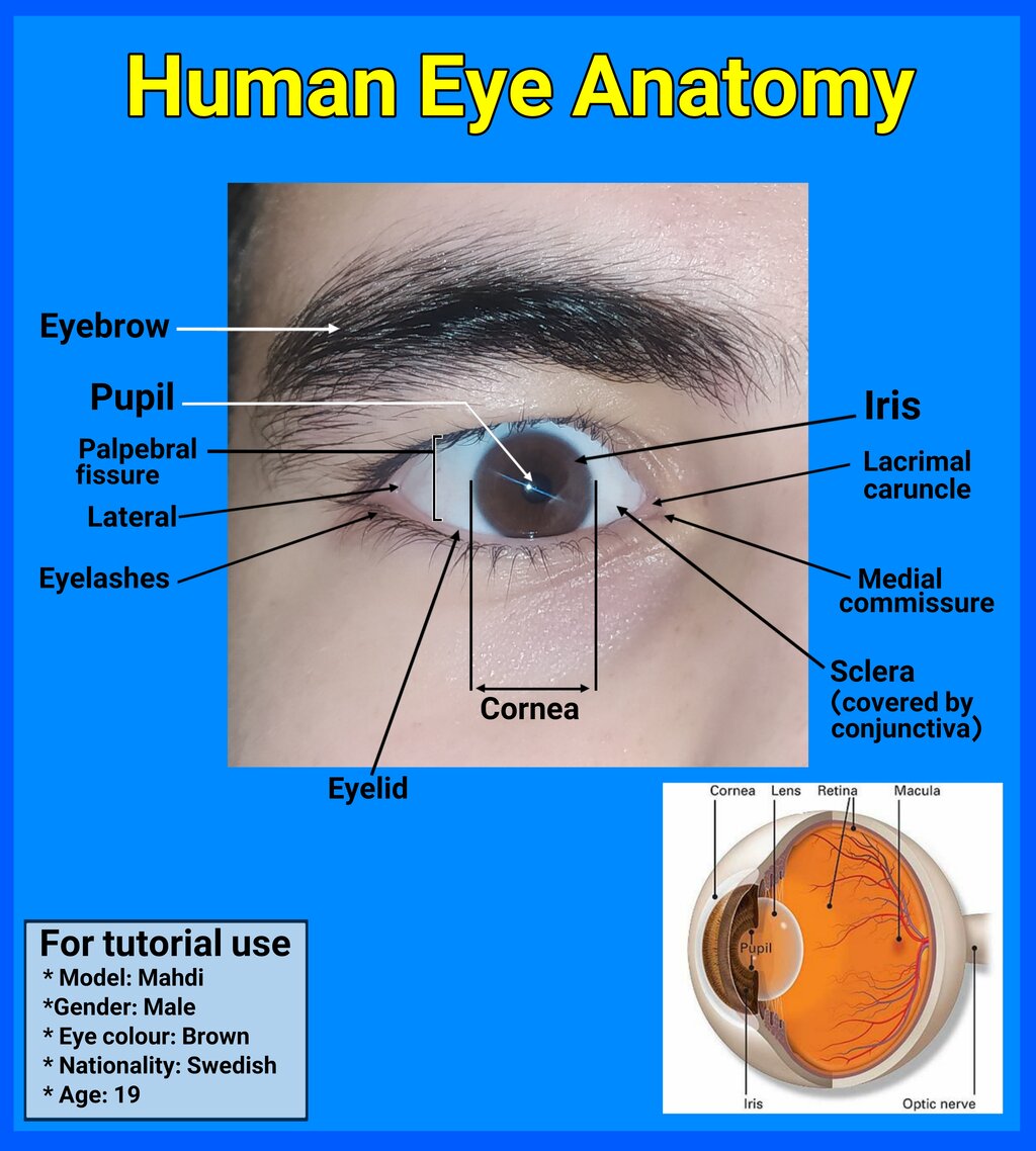
The journey of light begins at the front of the eye. The first thing it passes through is a transparent, dome-shaped layer.
Cornea
noun
The transparent outer layer at the very front of the eye that covers the iris, pupil, and lens. It helps focus incoming light.
Just behind the cornea is the colored part of the eye, the iris. The iris is a muscle that controls the size of the opening in its center, the pupil. By expanding and contracting, the iris regulates how much light enters the deeper parts of the eye. In bright light, the pupil gets smaller; in the dark, it opens up to let more light in.
Focusing the Picture
After passing through the pupil, light hits the lens. This structure is absolutely critical for clear vision, and it's where cataracts form.
The lens is a clear, flexible structure located directly behind the iris. Its main job is to fine-tune the focus of light, directing it precisely onto the back of the eye.
The eye is filled with two types of fluid that help it maintain its shape. The aqueous humor is a watery fluid that fills the space between the cornea and the lens. The vitreous humor is a thicker, gel-like substance that fills the large central cavity of the eye, behind the lens.
Creating the Image
Once light is focused by the cornea and lens, it travels through the vitreous humor to the back of the eye, where it lands on a light-sensitive layer of tissue.
Retina
noun
A thin layer of tissue at the back of the eye that contains millions of light-sensitive cells (rods and cones). The retina converts light into neural signals.
Think of the retina as the film in a camera or the sensor in a digital camera. It captures the focused light and begins the process of turning it into a coherent image.
Finally, the signals generated by the retina are sent to the brain through a bundle of nerve fibers.
The optic nerve acts like a data cable, transmitting visual information from the retina to the brain, which then interprets these signals as sight.
When all these parts work together, the result is clear vision. A cataract is a problem with just one of these parts: the lens. When the normally clear lens becomes cloudy, it can no longer focus light properly onto the retina, leading to blurry or obscured vision.
Let's review these key structures.
Now, check your understanding of the eye's anatomy.
What is the primary function of the iris?
A cataract is a clouding of which part of the eye?
With this foundation in place, you're ready to learn how a healthy lens becomes a cataract.