Bladder Ultrasound Fundamentals for Medical Professionals
Bladder Anatomy
Your Bladder's Place in the Pelvis
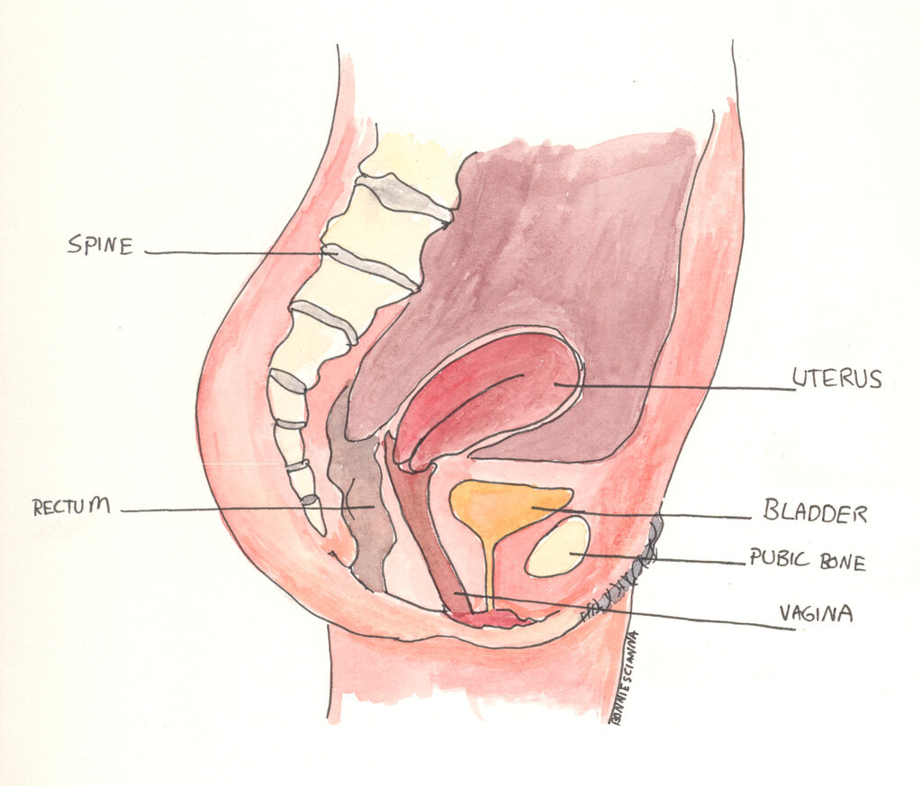
The urinary bladder is a hollow, muscular organ that sits low in your pelvis, right behind the pubic bone. Think of it as a flexible storage bag. When it's empty, it's about the size and shape of a pear. As it fills with urine from the kidneys, it expands into a rounder, more spherical shape.
Its location is slightly different depending on anatomy. In females, the bladder sits in front of the uterus. In males, it's located in front of the rectum and just above the prostate gland. This positioning is important because the bladder's neighbors can affect its function and how it appears on medical images.
The Bladder Wall
The bladder isn't just a simple sac. Its wall is made of several distinct layers, each with a specific job. This specialized structure is what allows it to stretch and contract so effectively.
The innermost lining is called the urothelium, a type of transitional epithelium. This tissue is remarkable for its ability to stretch as the bladder fills and then recoil to its original size after emptying, all without tearing. Beneath it are the lamina propria and submucosa, layers of connective tissue that contain blood vessels and nerves.
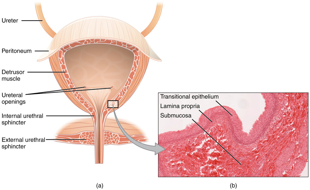
The most powerful layer is the detrusor muscle. This thick, smooth muscle forms the main body of the bladder. When it's time to urinate, the detrusor muscle contracts forcefully, squeezing the bladder to push urine out. The outermost layer is a fibrous covering that helps hold the bladder in place.
Storage and Voiding
The bladder has two main jobs: storing urine and then emptying it at the right time. This cycle of filling and voiding is a coordinated process involving muscles and nerves.
During the storage phase, the detrusor muscle is relaxed, allowing the bladder to expand and fill. At the same time, two sphincter muscles at the base of the bladder, where it connects to the urethra, remain tightly closed to prevent leaks.
As the bladder fills, stretch receptors in its wall send signals to your brain. When the volume reaches a certain point, you feel the urge to urinate. During voiding, also known as micturition, the process reverses. The brain signals the detrusor muscle to contract and the sphincter muscles to relax. This opens the passage, and the pressure from the contracting bladder pushes urine out of the body through the urethra.
Ready to test your knowledge of the bladder's anatomy and function?
What is the name of the innermost lining of the bladder, which is remarkable for its ability to stretch as the bladder fills?
In males, the bladder is located in front of the rectum and directly above which gland?
Understanding this basic anatomy is the first step in learning how to visualize the bladder with ultrasound.