Autonomic Nervous System Pharmacology
ANS Receptor Mechanics
The Machinery of the Message
When a neurotransmitter like norepinephrine or acetylcholine docks with a receptor, it's like a key turning in a lock. But what happens next? The signal doesn't just stop at the cell surface. It triggers a complex chain of events inside the cell, a process called signal transduction. This internal machinery determines whether a muscle contracts, a gland secretes, or a heart beats faster.
Most receptors in the autonomic nervous system don't act directly. Instead, they rely on intermediaries called G-proteins to relay the message. These proteins are like tiny molecular switches that, once activated, set off a specific cascade of chemical reactions.
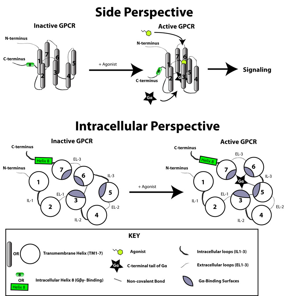
There are three main types of G-proteins we need to know for the ANS: Gs, Gi, and Gq. Think of them as different sub-routines a cell can run.
- Gs (stimulatory): Activates an enzyme called adenylyl cyclase, which increases the levels of a 'second messenger' molecule called cyclic AMP (cAMP).
- Gi (inhibitory): Does the opposite. It inhibits adenylyl cyclase, leading to a decrease in cAMP.
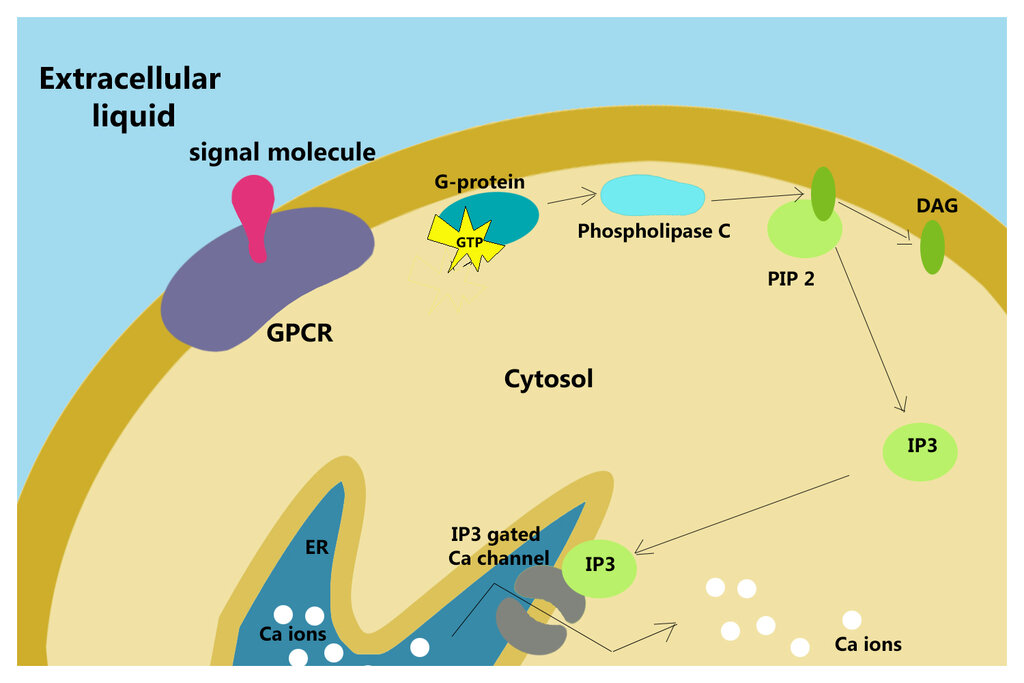
- Gq: Activates a different enzyme, phospholipase C. This triggers a pathway that increases intracellular calcium (Ca²⁺) levels, often leading to contraction or secretion.
Adrenergic Receptors
Adrenergic receptors respond to norepinephrine and epinephrine. Their effects depend entirely on which G-protein they're coupled to and where they are located in the body.
Alpha-1 (α1) receptors are coupled to Gq. When activated, they increase intracellular Ca²⁺. You find them primarily on the smooth muscle of blood vessels. The influx of calcium causes these muscles to contract, leading to vasoconstriction and an increase in blood pressure.
Alpha-2 (α2) receptors are linked to Gi. Their effect is inhibitory. A key location for α2 receptors is on the presynaptic nerve terminals. When norepinephrine is released into the synapse, some of it binds to these α2 receptors, creating a negative feedback loop that reduces further norepinephrine release. It's a self-regulating mechanism.
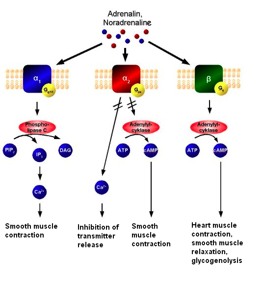
Now for the beta receptors. Both major types are coupled to Gs, so their general action is to increase cAMP. However, their locations lead to very different outcomes.
Beta-1 (β1) receptors are concentrated in the heart. Increased cAMP in cardiac muscle cells leads to a stronger, faster heartbeat. This increases cardiac output.
Beta-2 (β2) receptors are found on the smooth muscle of the bronchioles in the lungs and in certain blood vessels (like those supplying skeletal muscle). Here, increased cAMP has the opposite effect of α1 activation: it causes smooth muscle relaxation. This leads to bronchodilation (opening the airways) and vasodilation.
There are also Beta-3 (β3) receptors, primarily found in adipose tissue, which are also linked to Gs and play a role in lipolysis (the breakdown of fat).
Cholinergic Receptors
Cholinergic receptors bind acetylcholine (ACh) and are divided into two main families: muscarinic and nicotinic. Muscarinic receptors are G-protein-coupled, just like adrenergic receptors, while nicotinic receptors work very differently.
Muscarinic receptors are a family of five (M1-M5), but they follow a simple G-protein rule. A useful mnemonic is "QIQ".
- M1, M3, M5 receptors are coupled to Gq. Like α1 receptors, they increase intracellular Ca²⁺. M3 receptors are particularly important. They are found on smooth muscle (like in the gut and bladder) and in glands, where their activation promotes contraction and secretion, respectively.
- M2, M4 receptors are coupled to Gi. The classic example is the M2 receptor in the heart. Its activation decreases cAMP, which slows the heart rate.
Finally, we have . These are not GPCRs. They are ligand-gated ion channels. When two acetylcholine molecules bind to a nicotinic receptor, its central pore opens, allowing sodium ions (Na⁺) to rush into the cell. This rapid influx of positive charge causes immediate depolarisation.
This mechanism is built for speed. You find nicotinic receptors where fast transmission is essential: at the neuromuscular junction to trigger muscle contraction, and in all autonomic ganglia, where they transmit the signal from the pre-ganglionic to the post-ganglionic neuron.
| Receptor | G-Protein | Second Messenger | Key Effect & Location |
|---|---|---|---|
| α1 | Gq | ↑ IP₃, DAG, Ca²⁺ | Vasoconstriction (vascular smooth muscle) |
| α2 | Gi | ↓ cAMP | Inhibits neurotransmitter release (presynaptic) |
| β1 | Gs | ↑ cAMP | ↑ Heart rate & contractility (heart) |
| β2 | Gs | ↑ cAMP | Bronchodilation, vasodilation (lungs, muscle) |
| M1, M3, M5 | Gq | ↑ IP₃, DAG, Ca²⁺ | Secretion, contraction (glands, smooth muscle) |
| M2, M4 | Gi | ↓ cAMP | ↓ Heart rate (heart) |
| Nicotinic | None (Ion Channel) | Na⁺ influx | Depolarisation (autonomic ganglia, NMJ) |
Understanding this map of receptors, their signalling mechanisms, and their locations is the key to pharmacology. A drug that selectively blocks β1 receptors will slow the heart rate without affecting the airways. Conversely, a drug that stimulates β2 receptors can open up the lungs with minimal effect on the heart. The physiological outcome is all in the details of the receptor.