Architecting an All in One Medical Aesthetics EHR
Clinical Visual Documentation
Visual Charting with Coordinate Mapping
In aesthetics, precision is everything. Standard text notes like "injected 5 units into the forehead" are too vague for high-quality care. They lack the granular detail needed to replicate results, track subtle changes, or troubleshoot adverse effects. The solution is coordinate-based visual charting.
This system works by overlaying a digital map onto a high-resolution patient photograph. Instead of a freehand drawing, clinicians tap on specific points on the image. Each tap records a precise (x, y) coordinate. This coordinate becomes the anchor for all treatment data for that specific site.
For example, when treating glabellar lines, a clinician can tap three distinct points between the eyebrows. The system logs each point's coordinates and prompts for the details: product used, units injected, and lot number. This creates an unambiguous, data-rich record that's tied directly to the patient's unique anatomy.
This method transforms the patient's photo from a simple picture into an interactive clinical map. It serves as the foundation for everything that follows, from ensuring safety to visualizing progress.
The Camera-to-Chart Workflow
Clinical photos contain Protected Health Information (PHI) and require strict handling under HIPAA. The standard camera app on a mobile phone or tablet is a major compliance risk because it saves images to a local, often unencrypted, and cloud-synced photo gallery. This exposes sensitive data to potential breaches.
A secure 'camera-to-chart' workflow solves this by ensuring patient photos never touch the device's local storage. The process is seamless for the clinician but architecturally robust on the backend.
- Capture within the EHR: The clinician initiates the photo capture from within the secure, sandboxed EHR application.
- In-Memory Handling: The photo is captured directly into the application's memory, not the device's file system.
- Immediate Encryption: The image data is immediately encrypted using a strong algorithm (e.g., AES-256) before transmission.
- Direct Cloud Upload: The encrypted file is sent directly to a HIPAA-compliant cloud storage service, such as a private Amazon S3 bucket with server-side encryption enabled.
- No Local Trace: Once the upload is confirmed, the in-memory data is purged. No copy of the image ever resides in the device's public photo gallery.
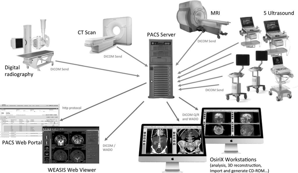
This workflow mitigates the risk of a lost or stolen device leading to a data breach. It treats every clinical photo with the same level of security as any other piece of the patient's electronic health record, because that's exactly what it is.
Data Structures and Progress Tracking
A visual chart is only as good as the data linked to it. The system's architecture must create a durable link between the (x, y) coordinates on an image and the specific treatment details. This is typically handled through a relational database where each injection point is a record in a treatments table.
This record links the patient, the specific photo used as a map, the coordinates, and all relevant medical metadata. Storing product lot numbers and expiration dates is not just good practice; it's a critical safety measure for tracking product batches in the event of a recall or adverse reaction.
{
  "imageId": "img_a7b3c9",
  "patientId": "p_112358",
  "timestamp": "2024-10-26T10:30:00Z",
  "annotations": [
    {
      "coordX": 450,
      "coordY": 812,
      "product": "Botox Cosmetic",
      "dosage": 5,
      "units": "units",
      "lotNumber": "C7139C3",
      "expiration": "2025-12-31"
    },
    {
      "coordX": 485,
      "coordY": 815,
      "product": "Botox Cosmetic",
      "dosage": 5,
      "units": "units",
      "lotNumber": "C7139C3",
      "expiration": "2025-12-31"
    }
  ]
}
This structured approach also powers two crucial features: before-and-after visualization and longitudinal tracking.
Before-and-after engines don't just place two photos side-by-side. A robust system uses facial recognition or manually placed anchor points (like the corners of the eyes and mouth) to align and scale the images correctly. This ensures a true, apples-to-apples comparison, correcting for slight changes in head tilt or distance from the camera.
Image versioning is essential for tracking progress over years. Each new photo is another version in the patient's visual history. This allows a clinician to see not just the last visit's results, but the cumulative effect of treatments over time. By linking each photo back to the detailed treatment data, a clinician can analyze which techniques and dosages delivered the best long-term outcomes, enabling truly personalized and data-driven aesthetic care.
Clinical photographs have the ability to capture a rich amount of data in an image, forming an integral part of consultation, medical education, and publication in plastic surgery.
By combining precise mapping, secure workflows, and linked data, we transform clinical photos from static images into dynamic, queryable records that enhance patient safety and treatment efficacy.
What is the primary limitation of standard text-based notes (e.g., "injected 5 units into the forehead") in aesthetics?
Why is using a standard mobile device camera app to take patient photos a major compliance risk?