Anatomy of the Human Skull
Neurocranium Architecture
The Cranial Vault
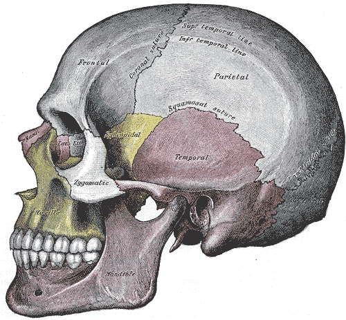
The neurocranium forms the protective case around the brain. It's composed of eight distinct bones: the unpaired frontal, occipital, sphenoid, and ethmoid bones, along with the paired parietal and temporal bones. These aren't just fused together haphazardly; they meet at fibrous joints called sutures, creating a strong but slightly flexible dome.
We can divide the neurocranium into two main parts: the calvaria (the skullcap or vault) and the cranial base (the floor). The vault is the smooth, curved upper portion you typically think of as the skull. Its primary job is to absorb and distribute the force of impacts.
The bones of the vault have a unique three-layered structure, much like a sandwich. The outer and inner layers are made of dense compact bone, called the external and internal tables. Between them is a layer of spongy bone called the diploë which contains red bone marrow.
This structure is an evolutionary marvel. The tough outer table resists fracture, while the spongy diploë can compress and absorb shock, protecting the brittle inner table from shattering and damaging the delicate brain tissue beneath.
Key Bones and Junctions
Let's focus on two of the major players in the cranial vault: the frontal and parietal bones.
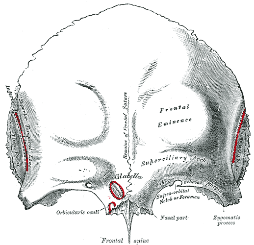
The frontal bone forms the forehead and the superior part of the orbits (eye sockets). It houses the frontal sinuses, which are air-filled cavities that lighten the skull and contribute to voice resonance. These sinuses drain into the nasal cavity. Above the orbits, you can feel two arches, the superciliary arches, which are more prominent in males.
The two parietal bones form the bulk of the sides and roof of the cranium. They are large, quadrilateral bones that articulate with each other at the sagittal suture, with the frontal bone at the coronal suture, and with the occipital bone at the lambdoid suture.
Where these major bones meet, specific landmarks are formed. The most clinically significant junction is the pterion a relatively weak H-shaped point on the side of the skull where the frontal, parietal, temporal, and sphenoid bones meet.
Just deep to the pterion runs the middle meningeal artery. A fracture at this point can rupture the artery, leading to an epidural hematoma, a life-threatening condition where blood collects between the dura mater and the skull, compressing the brain. This is why a blow to the temple is so dangerous.
From Membrane to Bone
Unlike the long bones of your limbs, which form from cartilage models, the flat bones of the cranial vault develop through a process called intramembranous ossification This process begins in the embryo when mesenchymal cells, a type of stem cell, differentiate directly into bone-producing cells (osteoblasts).
These osteoblasts secrete an organic matrix called osteoid, which then calcifies. This happens at multiple points called ossification centers, and bone tissue radiates outward from these centers. As the developing brain expands, these bony plates grow larger until they meet to form the sutures. This direct-to-bone formation allows for the rapid growth needed to enclose the brain.
Calvaria
noun
The upper, dome-like part of the skull that encloses the brain, also known as the skullcap or cranial vault.
Variations in this ossification process, especially the premature fusion of sutures (craniosynostosis), can significantly alter the shape of the head and restrict brain growth. The specific shape depends on which suture fuses too early. For instance, premature fusion of the sagittal suture results in a long, narrow skull, while fusion of the coronal suture can lead to a flattened, wide skull.
Which of the following bones of the neurocranium is unpaired?
What is the primary function of the diploë, the spongy layer found between the inner and outer tables of the skull's vault?
Understanding the architecture of the neurocranium is fundamental to appreciating how the brain is both protected and accommodated as it grows.