Anatomy and Clinical Significance of the Popliteal Fossa
Popliteal Fossa Boundaries
The Popliteal Fossa
Behind your knee is a shallow, diamond-shaped depression known as the popliteal fossa. It’s more than just the back of the knee; it's a critical passageway for major nerves and blood vessels travelling between the thigh and the lower leg. Understanding its precise boundaries is key to locating structures and diagnosing issues in this region.
Muscular Borders
The popliteal fossa is defined not by bones, but by the arrangement of powerful muscles from the thigh and leg.
Two of the hamstring muscles, the semimembranosus and the semitendinosus, travel down the back of the thigh to form the upper medial (superomedial) border. On the other side, the biceps femoris muscle, another hamstring, forms the upper lateral (superolateral) border.
Below the knee, the two heads of the large calf muscle, the gastrocnemius, create the lower boundaries. The medial head forms the inferomedial border, and the lateral head forms the inferolateral border.
| Boundary | Muscle(s) |
|---|---|
| Superomedial | Semimembranosus & Semitendinosus |
| Superolateral | Biceps femoris |
| Inferomedial | Medial head of gastrocnemius |
| Inferolateral | Lateral head of gastrocnemius |
Floor and Roof
The fossa isn't an empty space; it has a floor beneath it and a roof covering it.
The floor, or the deep surface, is formed by several structures. From top to bottom, these are the popliteal surface of the femur, the posterior capsule of the knee joint, and a strong fascia covering the popliteus muscle.
The roof is much simpler. It consists of the tough popliteal fascia lying just under the skin. This fascia is continuous with the fascia lata of the thigh and the deep fascia of the leg, creating a strong, containing layer over the fossa's contents.
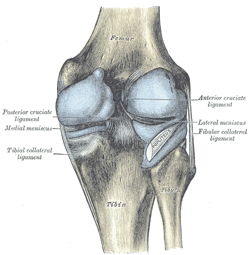
These precise muscular and fascial boundaries are clinically important. They act like a container. Any swelling, such as from a Baker's cyst (a fluid-filled sac), infection, or aneurysm of the popliteal artery, will be confined within this space. Knowing the borders helps predict how and where this pressure might spread, guiding diagnosis and treatment.
Now, let's test your understanding of this important anatomical space.
Which muscle(s) form the superomedial border of the popliteal fossa?
A patient presents with a swelling behind their knee, which is confined to a diamond-shaped area. The defined boundaries of the popliteal fossa are clinically important because they: