Anatomy and Clinical Assessment of the Anterior Thigh
Anterior Compartment Architecture
The Five-Headed Quadriceps
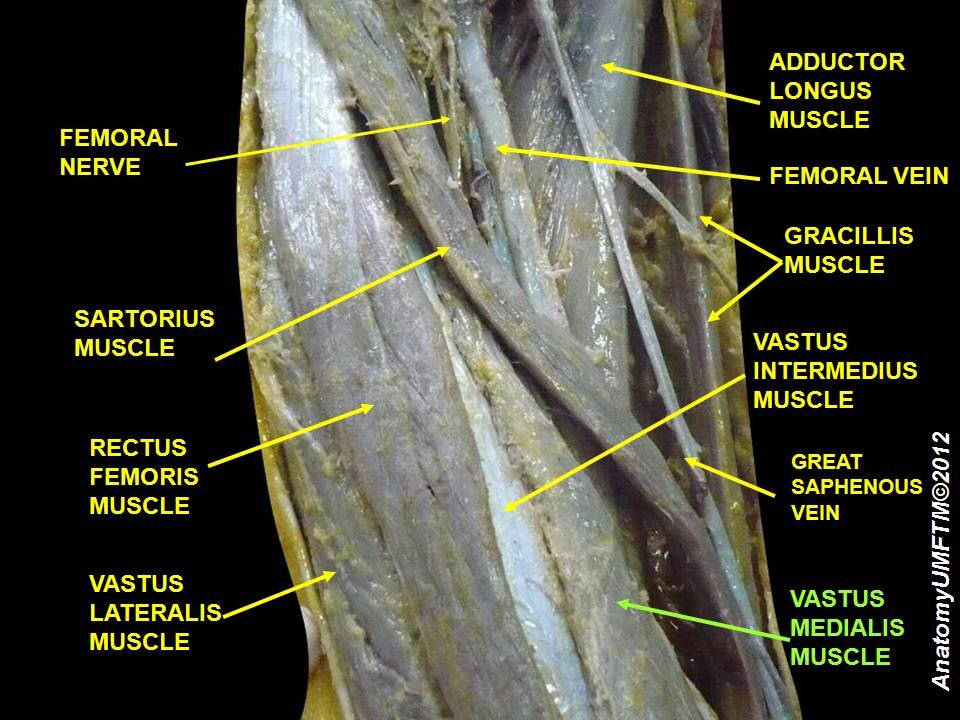
The anterior compartment of the thigh is dominated by a powerful group of muscles collectively known as the quadriceps femoris. Traditionally, this group is described as having four heads: the rectus femoris, vastus lateralis, vastus medialis, and vastus intermedius. However, recent anatomical studies have identified a fifth functional component: the tensor of the vastus intermedius (TVI).
This isn't just an academic detail. The TVI has its own distinct aponeurosis, a sheet-like tendon, that merges with the aponeuroses of the other vasti muscles. Its primary role appears to be tensioning the vastus intermedius and pulling on the quadriceps tendon from a unique angle. Understanding this five-part structure is crucial for diagnosing and treating knee injuries, especially those involving the extensor mechanism.
The four muscles collectively insert onto the patella via the quadriceps tendon.
Each head has a specific origin. The vasti muscles (lateralis, medialis, and intermedius) are mono-articular, meaning they only cross one joint—the knee. They originate from various points along the shaft of the femur. In contrast, the rectus femoris is bi-articular; it crosses both the hip and the knee. It originates from the anterior inferior iliac spine of the pelvis. This dual-joint action allows it to not only extend the knee but also to flex the hip, a function essential for actions like kicking or sprinting.
The different lines of pull from each quadriceps head, including the TVI, are critical for patellar tracking—how the kneecap moves within its groove on the femur. An imbalance, often weakness in the vastus medialis, can lead to improper tracking and anterior knee pain. The distinct aponeuroses of the vasti muscles create a layered, complex structure. These fibrous sheets don't just transmit force; they allow for subtle, independent contractions that fine-tune knee movement.
Key Players and Their Roles
While the quadriceps are the main engine of the anterior thigh, two other muscles play vital roles: the sartorius and the iliopsoas.
The sartorius is the longest muscle in the human body, crossing both the hip and knee joints. It's a thin, strap-like muscle that runs obliquely across the thigh, from the anterior superior iliac spine to the medial side of the tibia. Its unique path allows it to flex, abduct, and laterally rotate the hip, as well as flex the knee. Think of the motion of crossing one leg over the other while sitting—that's the sartorius at work.
The iliopsoas is often considered the primary hip flexor. It's a deep composite muscle formed by the psoas major and the iliacus. While its main muscle bellies are in the posterior abdomen and pelvis, its tendon crosses the hip joint anteriorly to insert on the lesser trochanter of the femur. In the thigh, it passes deep to the inguinal ligament and forms the floor of a region called the femoral triangle. Tightness or inflammation of the iliopsoas tendon is a common source of anterior hip pain, particularly in athletes who perform repetitive hip flexion.
Clinical Significance
The anatomy of the anterior compartment has direct implications for sports medicine. The quadriceps are responsible for decelerating the body, like when landing from a jump, which places immense eccentric load on the extensor mechanism. Ruptures of the quadriceps tendon or patellar tendon are severe injuries that can result from this forceful loading.
Furthermore, the rectus femoris, as a bi-articular muscle, is particularly susceptible to strain injuries, especially at its proximal origin. Understanding the layered aponeurotic connections of the vasti and TVI is also changing how surgeons approach knee surgery and how physiotherapists design rehabilitation programmes to restore balanced muscle function and proper patellar tracking.
Time to test your knowledge of the anterior thigh's architecture.
Recent anatomical studies have identified a fifth functional component of the quadriceps femoris group. What is this muscle called?
Which of the following muscles is considered 'bi-articular', meaning it crosses both the hip and the knee joints?
By understanding the individual roles and coordinated function of these muscles, we can better appreciate the complex mechanics of hip and knee movement.