Advancing Alzheimers Understanding and Care
Molecular Pathophysiology Mechanisms
The Protein Villains
At the heart of Alzheimer's disease are two misbehaving proteins: amyloid-beta and tau. For a long time, the leading theory, known as the amyloid cascade hypothesis, has placed the primary blame on amyloid-beta. This peptide is a fragment of a larger protein called Amyloid Precursor Protein (APP), which is embedded in the membrane of neurons. In healthy brains, APP is cleaved by enzymes in a way that produces harmless fragments that are easily cleared away.
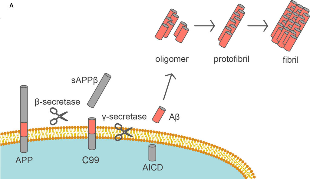
In Alzheimer's, this process goes wrong. Two different enzymes, beta-secretase and gamma-secretase, cut APP at the wrong spots. This releases a sticky, 42-amino-acid-long fragment called amyloid-beta 42 (Aβ42). Unlike its shorter, less problematic relatives, Aβ42 has a strong tendency to misfold and clump together. First, individual monomers form small, soluble clusters called oligomers. These oligomers are now considered the most neurotoxic form of amyloid-beta, capable of directly interfering with synaptic function long before they form larger structures. Over time, these oligomers aggregate further into fibrils and eventually deposit as large, insoluble extracellular plaques between neurons.
These amyloid plaques act like disruptive boulders in the brain, physically obstructing communication between neurons and triggering a destructive inflammatory response.
An Internal Collapse
While amyloid plaques build up outside the neurons, a different kind of chaos unfolds within. This involves the tau protein. Normally, tau acts as a stabilizing support for microtubules, which are like the internal railway tracks of a neuron. These tracks are essential for transporting nutrients, organelles, and neurotransmitters from the cell body down the axon.
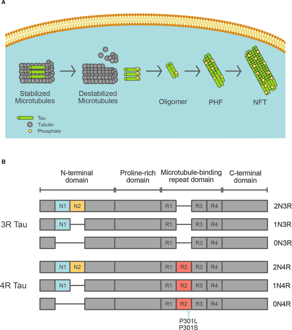
In Alzheimer's, tau undergoes a chemical change called hyperphosphorylation, where an excessive number of phosphate groups attach to it. This causes the tau protein to change shape, detach from the microtubules, and lose its ability to stabilize them. The transport system breaks down, and the neuron's internal structure collapses. These detached, misfolded tau proteins then clump together inside the cell, forming insoluble twisted fibers known as neurofibrillary tangles (NFTs). The neuron, now choked with tangles and with a defunct transport system, eventually dies.
A crucial part of the story is how these two pathologies connect. Evidence suggests a 'dual pathway' where the extracellular amyloid-beta oligomers and plaques somehow trigger or accelerate the intracellular tau hyperphosphorylation. The exact mechanism is still under investigation, but it appears the toxic environment created by amyloid stress puts neurons under pressure, activating kinases (enzymes that add phosphate groups) that go on to modify tau. This creates a devastating one-two punch: disruption from the outside and collapse from within, leading to widespread synaptic dysfunction and neuronal apoptosis (programmed cell death).
Risk Factors and Inflammation
Why do these proteins go rogue in some people but not others? Genetics plays a significant role, most notably with the apolipoprotein E (APOE) gene. This gene comes in three common forms, or alleles: APOE-ε2, APOE-ε3, and APOE-ε4. The APOE-ε4 allele is the strongest known genetic risk factor for late-onset Alzheimer's disease. Individuals with one copy of APOE-ε4 have a significantly increased risk, and those with two copies have an even higher risk. The APOE protein is involved in transporting cholesterol and clearing fats, but in the brain, it's also believed to play a role in clearing amyloid-beta. The ε4 version seems to be less efficient at this cleanup job, allowing Aβ42 to accumulate more easily.
The brain doesn't sit idly by as these plaques and tangles form. The presence of amyloid plaques triggers a chronic inflammatory response. The brain's resident immune cells, called microglia, become activated. In the short term, this is a protective response, as microglia attempt to clear the plaques through phagocytosis (engulfing and digesting debris). However, in a state of chronic activation, these microglia can become dysfunctional. They release inflammatory molecules like cytokines and chemokines, which, while intended to fight pathogens, can cause collateral damage to surrounding neurons. This state of persistent contributes to synaptic dysfunction and exacerbates the neurodegenerative process, creating a vicious cycle where inflammation drives further pathology.
According to the amyloid cascade hypothesis, which of the following is considered the initial event that leads to the pathological changes seen in Alzheimer's disease?
What is the normal function of the tau protein in a healthy neuron?
Understanding these molecular drivers—amyloid plaques, tau tangles, genetic predispositions, and chronic inflammation—is key to grasping the biological reality of Alzheimer's disease and the rationale behind modern therapeutic strategies.