Advanced Thoracic Sympathetic Trunk Anatomy
Segmental Organization
The Sympathetic Chain's Bony Ladder
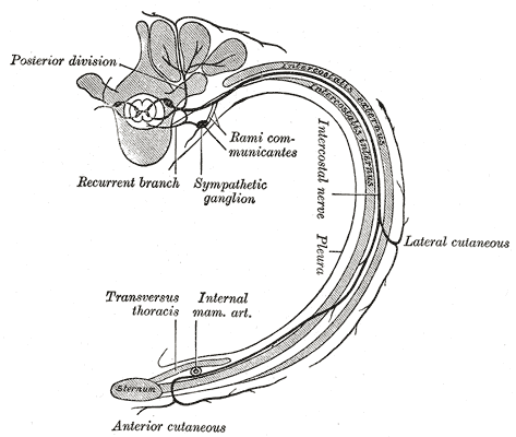
The sympathetic chain runs parallel to the vertebral column, like a ladder of nerves on each side. The "rungs" of this ladder are the paravertebral ganglia, which are clusters of nerve cell bodies. In the thorax, these ganglia are anchored against the heads of the ribs, right where they connect to the vertebrae at the costovertebral joints. This fixed position provides a reliable anatomical landmark.
Typically, there are 11 or 12 thoracic ganglia on each side, but this number isn't always rigid. Sometimes, ganglia fuse together during development. The entire chain sits in a specific compartment, tucked behind the parietal pleura, the membrane lining the chest cavity. This retropleural position means it's outside the space occupied by the lungs, safely separated from their movements.
The Stellate Ganglion A Special Case
At the top of the thoracic chain, a significant anatomical variation occurs. The inferior cervical ganglion often fuses with the first thoracic ganglion. This creates a larger, star-shaped structure known as the cervicothoracic ganglion, or more commonly, the stellate ganglion due to its radiating nerve processes. It is typically found anterior to the neck of the first rib and extends up toward the C7 vertebra.
This fusion is clinically important. Because it consolidates sympathetic innervation for the head, neck, arm, and upper chest, the stellate ganglion is a key target for therapeutic nerve blocks. This single, accessible point can be used to treat a range of conditions, from chronic pain syndromes in the arm to circulatory issues.
On-Ramps and Off-Ramps
To understand how the sympathetic chain functions, think of it as a highway running alongside the spinal cord. Spinal nerves need on-ramps and off-ramps to communicate with it. These ramps are the rami communicantes.
The white rami communicantes are the on-ramps. They carry preganglionic nerve fibers from the spinal cord to the sympathetic chain. These fibers are myelinated, which gives them a whitish appearance. Crucially, these on-ramps only exist from the T1 to L2 spinal levels. This is the entire origin point for the body's sympathetic outflow.
The gray rami communicantes are the off-ramps. They carry postganglionic fibers from the sympathetic chain back to the spinal nerves, which then distribute them to the body. These fibers are unmyelinated, giving them a grayish look. Unlike the white rami, every spinal nerve has a gray ramus. This allows sympathetic signals that originated in the T1-L2 region to be distributed to every part of the body, from head to toe.
| Ramus Type | Function | Myelination | Spinal Levels |
|---|---|---|---|
| White Ramus Communicans | Carries preganglionic fibers TO chain | Myelinated | T1 – L2/L3 only |
| Gray Ramus Communicans | Carries postganglionic fibers FROM chain | Unmyelinated | All 31 spinal levels |
The segmental organization of this system has profound surgical implications. Because the inputs (white rami) are limited to the thoracic and upper lumbar regions, surgeons can perform highly targeted procedures. A thoracic sympathectomy, for example, involves cutting or clamping the chain at specific levels to treat conditions like hyperhidrosis (excessive sweating) in the hands or face. Knowing the precise location of the ganglia relative to the ribs allows surgeons to access the chain with minimally invasive techniques, disrupting only the necessary segments while preserving function elsewhere.
The white rami communicantes serve as 'on-ramps' to the sympathetic chain. What is a key characteristic of these structures?
The stellate ganglion is a clinically significant structure formed by the fusion of which two ganglia?