Advanced Spinal Stenosis Management
Advanced Diagnostic Techniques
Peeking Inside the Spine
When symptoms suggest spinal stenosis, doctors need to see exactly what's happening inside the spinal canal. While a standard X-ray can show the alignment of your vertebrae, it doesn't reveal the soft tissues. That's where more advanced imaging comes in.
Magnetic Resonance Imaging, or MRI, is the go-to tool for this job. It uses powerful magnets and radio waves to create detailed, cross-sectional images of the spine. Think of it like looking at individual slices of a loaf of bread. This technique excels at showing soft tissues like the spinal cord, nerves, and intervertebral discs.
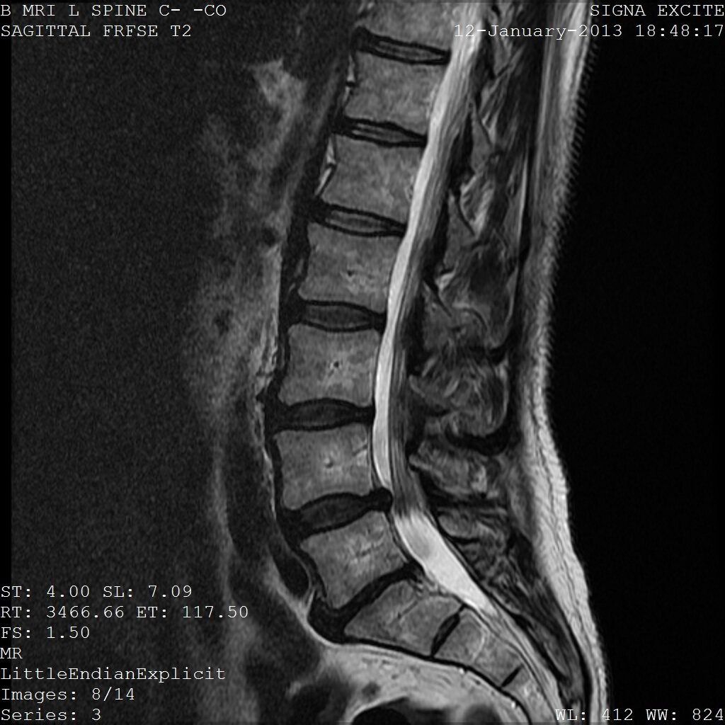
On an MRI, a radiologist can see if a bulging disc or thickened ligament is pressing on a nerve root or the spinal cord itself. The images clearly show the degree of narrowing in the spinal canal and the foramina—the small openings where nerves exit the spine. This information is crucial for pinpointing the exact location and severity of the stenosis.
A Closer Look at Bone
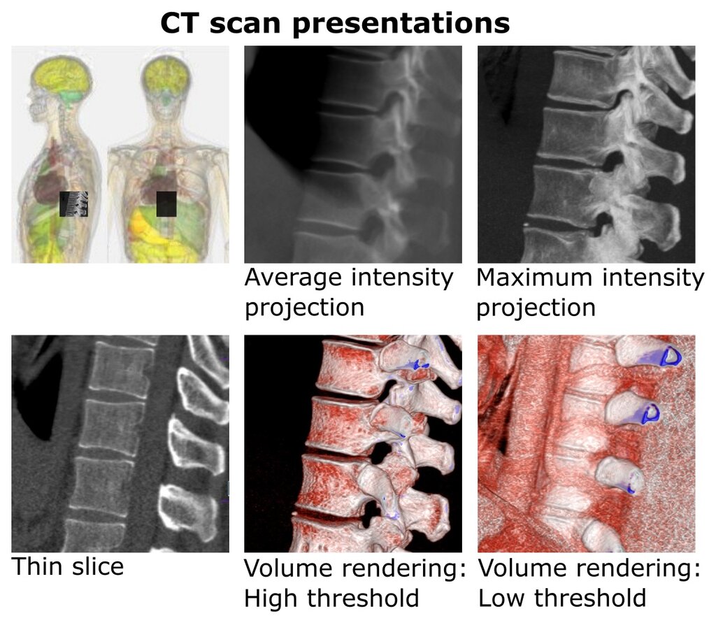
Sometimes, the problem isn't just with soft tissues. Computed Tomography, or a CT scan, provides a different kind of insight. A CT scan is essentially a sophisticated, 3D X-ray that creates detailed images of your bones.
This method is excellent for visualizing bony structures. It can clearly identify bone spurs (osteophytes), fractures, or arthritis in the facet joints that might be contributing to the narrowing of the spinal canal. It provides a precise map of the bony anatomy, which is vital for surgical planning.
For an even clearer picture of how the nerves are affected, a doctor might order a CT myelogram. In this procedure, a special dye is injected into the spinal fluid. The dye acts as a contrast agent, making the spinal cord and nerve roots appear bright white on the subsequent CT scan. This clearly outlines any areas where the flow of spinal fluid is being blocked by a herniated disc or bone spur.
A myelogram is particularly useful for patients who can't have an MRI, perhaps because they have a pacemaker or certain metal implants. It provides a dynamic look at the spine and the space available for the nerves.
Testing the Wires
Imaging tests show the spine's structure, but they don't tell the whole story. They can reveal that a nerve is being compressed, but not necessarily how that nerve is functioning. To assess nerve health directly, doctors use tests that measure electrical activity.
An Electromyography (EMG) test evaluates the health of muscles and the nerve cells that control them. Small needles are inserted into specific muscles to record their electrical activity, both at rest and during contraction. If a nerve is damaged from compression, the muscle's electrical signals might be abnormal.
A Nerve Conduction Study (NCS) is often done alongside an EMG. This test measures how fast an electrical impulse moves through your nerve. Electrodes are placed on the skin over the nerve, and a small shock is used to stimulate it. By measuring the speed of the signal, doctors can determine if there's nerve damage.
Think of it this way: an MRI or CT is like looking at a highway for traffic jams. An EMG and NCS is like checking the speed of the cars on that highway to see if they're actually slowing down.
Together, these tests help confirm if the anatomical narrowing seen on an imaging scan is the true source of a patient's symptoms, like weakness or numbness. They distinguish nerve damage from other conditions and help guide treatment decisions.
The Future of Diagnostics
The field of spinal diagnostics is constantly evolving, with new technologies promising even greater accuracy and efficiency.
AI-assisted imaging analysis is one of the most exciting developments. Artificial intelligence algorithms can be trained to analyze MRI and CT scans, quickly identifying and measuring areas of stenosis that might be subtle to the human eye. This can help standardize the interpretation of scans and reduce the time it takes to get a diagnosis.
Robotic-ultrasound navigation systems are another emerging tool. These systems combine real-time ultrasound imaging with robotics to create detailed 3D models of the spine without any radiation. This technology can help guide procedures and provide a dynamic view of how movement affects the spinal canal.
These advanced tools are paving the way for more personalized and precise treatments for spinal stenosis, helping doctors not only see the problem but also understand its functional impact.
What is the primary advantage of using an MRI to diagnose spinal stenosis?
A patient with a pacemaker cannot undergo an MRI. Which imaging test is a suitable alternative for visualizing potential nerve compression with high detail?
Each diagnostic tool offers a unique window into the complexities of the spine, helping build a complete picture for an accurate diagnosis.