Advanced Psychobiotics and Neurometabolism
Vagal Signaling Architecture
Neuropod Cells as Vagal Transducers
The rapid transmission of gut sensory information to the brain, occurring in milliseconds, cannot be explained by slow-acting hormonal pathways alone. This speed points to a direct neural connection, a role fulfilled by a specialized subset of enteroendocrine cells known as neuropod cellss. These cells act as primary sensory transducers, forming bona fide synapses directly with vagal afferent nerve endings. This arrangement creates a neuroepithelial circuit that bypasses slower humoral signaling.
Upon stimulation by luminal contents, such as sugars or bitter compounds, neuropod cells depolarize. This triggers the release of glutamate from synaptic vesicles in a quantal fashion, directly activating postsynaptic receptors on vagal afferents. The kinetics of this glutamatergic release are remarkably fast, enabling near-instantaneous communication of nutrient presence. This mechanism is distinct from the slower, more diffuse release of peptides like cholecystokinin (CCK), which also originates from intestinal chemosensory cells but acts over longer timescales and distances.
Microbial Metabolites and Vagal Firing
The gut microbiota doesn't just influence the host through immune modulation; it directly modulates vagal signaling. Bacterial metabolites, particularly short-chain fatty acids (SCFAs) like butyrate, acetate, and propionate, serve as key signaling molecules. These SCFAs can directly activate vagal afferents by binding to specific G-protein coupled receptors, such as FFAR2 and FFAR3, expressed on the nerve terminals themselves. This interaction triggers downstream signaling cascades that alter neuronal excitability.
This direct chemical-to-electrical transduction allows the brain to monitor the metabolic state of the gut microbiota in real time. Different ratios of SCFAs can produce distinct patterns of vagal firing, encoding information about microbial composition and activity.
The afferent signals generated by these metabolites travel up the vagus nerve to the (NTS) in the brainstem. The NTS acts as a primary integration center for visceral sensory information. The firing rate and pattern of NTS neurons are directly modulated by the incoming vagal input, effectively translating the gut's microbial signature into a neural code. Specific bacterial ligands can thereby selectively increase or decrease the spontaneous firing rates of NTS neurons.
Modulating Emotion via NTS-Amygdala Circuits
From the NTS, visceral information is relayed to higher brain centers, including the limbic system. A key pathway involves projections from the NTS to the central nucleus of the amygdala (CeA), a critical region for processing fear, anxiety, and emotional salience. The modulation of NTS firing rates by gut-derived signals therefore directly influences amygdalar activity.
For instance, specific probiotic strains capable of producing anti-inflammatory metabolites can increase vagal tone. This leads to a distinct signaling pattern in the NTS, which subsequently dampens hyperactivity in the CeA, producing an anxiolytic effect. Conversely, dysbiotic states can generate pro-inflammatory signals that are transduced into neural activity patterns associated with heightened anxiety and stress responses. This circuitry provides a direct neuroanatomical basis for the well-documented link between gut health and mood.
Paracrine Versus Endocrine Signaling
The communication strategy of enteroendocrine cells is not monolithic. While neuropod cells engage in direct, rapid synaptic transmission, other EECs utilize slower signaling modalities. The key distinction lies between paracrine and endocrine action.
In paracrine signaling, an EEC releases signaling molecules, such as serotonin (5-HT) or CCK, that diffuse locally to act on nearby cells, including adjacent epithelial cells or submucosal nerve endings. This is a neighborhood-level communication system, faster than hormonal signaling but more diffuse than synaptic transmission.
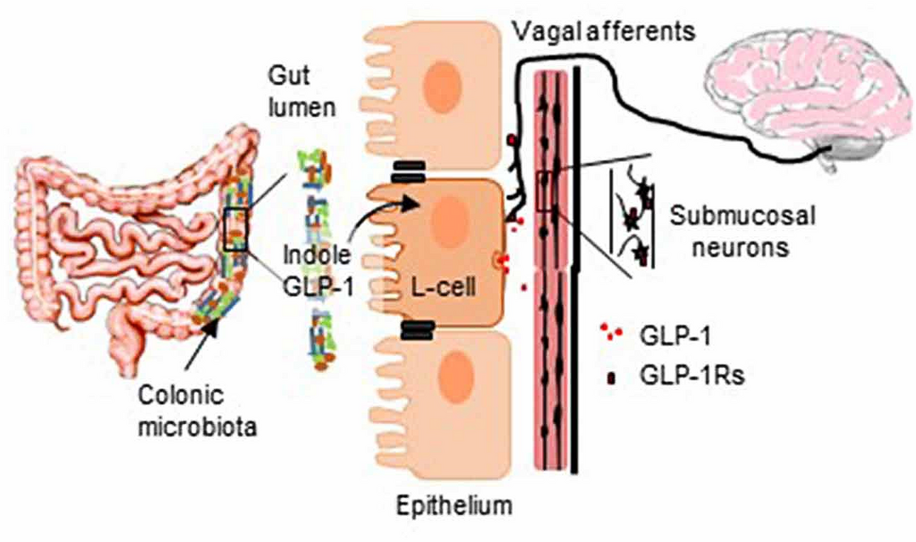
Endocrine signaling, by contrast, involves the release of hormones like GLP-1 or PYY into the bloodstream. These molecules travel systemically to act on distant targets, including the pancreas, adipose tissue, and the brain itself, particularly in areas lacking a robust blood-brain barrier like the hypothalamus. This pathway governs long-term metabolic regulation and satiety over minutes to hours.
The gut therefore employs a multi-layered communication strategy: fast synaptic signaling for immediate luminal updates, local paracrine networks for regional coordination, and slow endocrine signals for systemic metabolic control.
What is the primary mechanism that allows neuropod cells to transmit sensory information from the gut to the brain in milliseconds?
Which of the following best describes the role of the Nucleus Tractus Solitarius (NTS) in the gut-brain axis?
This architecture highlights a sophisticated system where the nervous system directly eavesdrops on the gut's chemical environment, enabling a rapid and nuanced gut-brain dialogue.