Advanced Neurology Exploration
Advanced Neuroimaging Techniques
Watching the Brain at Work
While structural scans give us a static picture of the brain's anatomy, functional Magnetic Resonance Imaging (fMRI) provides a movie. It tracks brain activity in real time by measuring changes in blood flow. When a brain area becomes more active, it needs more oxygen. The fMRI machine detects the magnetic properties of oxygenated and deoxygenated blood, creating a dynamic map of brain function.
This process relies on the Blood-Oxygen-Level-Dependent (BOLD) signal. More active neurons demand more oxygen-rich blood, and this change in blood oxygenation is what the fMRI scanner picks up.
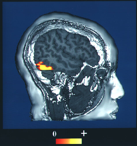
Researchers use fMRI to see which parts of the brain light up when a person performs a task, thinks about something, or experiences an emotion. It helps us understand the neural basis for everything from language to memory. But fMRI is just one tool for looking at brain function. To see the brain's metabolic processes, we need a different approach.
Tracing Metabolic Activity
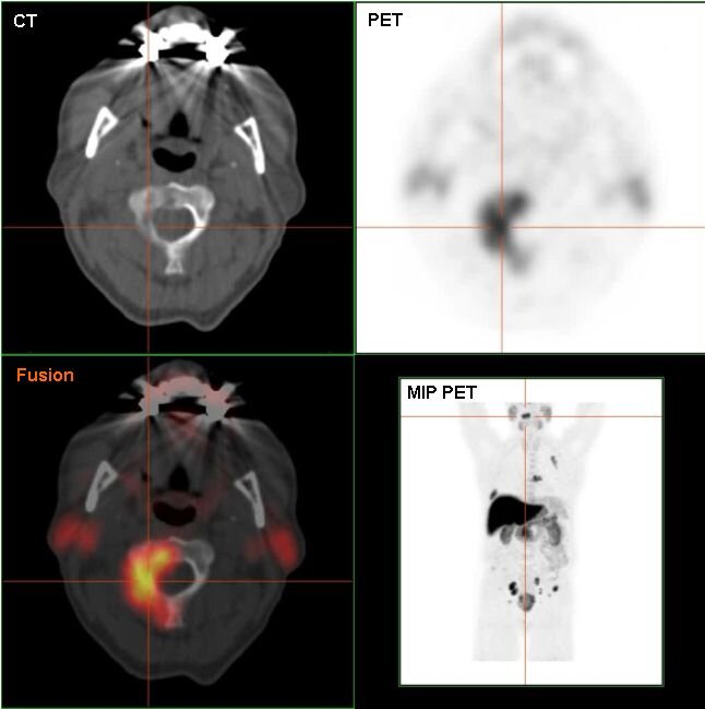
Positron Emission Tomography (PET) scans offer a window into the brain's chemical activity. Before a PET scan, a patient receives an injection of a small amount of a radioactive tracer. This tracer is attached to a natural substance, like glucose, which the brain uses for energy. The scanner then tracks where this tracer accumulates.
Areas with high metabolic activity, such as rapidly growing tumors, will use more glucose and show up as bright spots on the PET image. This technique is also invaluable for diagnosing neurodegenerative diseases. For example, specific tracers can be designed to bind to amyloid plaques, which are a hallmark of Alzheimer's disease, making them visible on a scan long before severe symptoms appear.
AI and the Future of Imaging
Neuroimaging techniques like fMRI and PET produce enormous amounts of complex data. A single scan can contain millions of data points, making manual analysis challenging and time-consuming. This is where artificial intelligence (AI) and machine learning (ML) come in.
AI algorithms can sift through these massive datasets to identify subtle patterns that might be invisible to the human eye. They can help differentiate between healthy and diseased tissue with greater accuracy, predict how a patient might respond to a particular treatment, and even help diagnose conditions like autism or depression based on functional brain patterns.
The rise of deep learning algorithms, such as convolutional neural networks (CNNs), offers fascinating perspectives for the automation of medical image analysis.
By integrating AI with neuroimaging, clinicians can make more precise diagnoses and researchers can gain deeper insights into the complex workings of the brain. This fusion of technologies is pushing the boundaries of what we can learn about neurological health and disease.
Ready to test your knowledge?