Advanced Neural Systems and Cognitive Function
Synaptic Modulation Mechanisms
Beyond the Ion Channel
Fast, ionotropic responses are the nervous system's workhorses. An action potential arrives, glutamate binds to an AMPA receptor, a channel opens, and sodium ions rush in. It's a direct, reliable way to pass a signal. But this isn't the whole story. For the brain to learn and adapt, it needs a more nuanced, longer-lasting form of communication. This is the world of synaptic modulation, driven by metabotropic receptors.
Instead of being ion channels themselves, metabotropic receptors initiate a cascade of intracellular events. Think of an ionotropic receptor as a simple light switch: on or off. A metabotropic receptor is more like a smart home hub. It receives a signal and can then dim the lights, adjust the thermostat, and start a playlist, all from a single command. The most common type of these
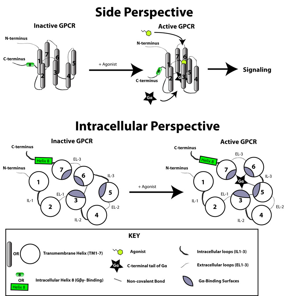
smart hubs are G-protein coupled receptors (GPCRs). When a neurotransmitter binds to a GPCR, it doesn't open a channel directly. Instead, it activates an associated G-protein, which then kicks off a chain reaction inside the cell. This process is slower, but its effects are far more widespread and enduring.
The Second Messengers
Once a G-protein is activated, it often stimulates an enzyme to produce intracellular signaling molecules called second messengers. The neurotransmitter is the "first messenger," delivering the signal to the cell's outer membrane. The second messengers take that signal and broadcast it throughout the cell's interior, amplifying it along the way.
Two major second messenger pathways are crucial for synaptic modulation:
-
The cAMP Pathway: The G-protein activates an enzyme called adenylyl cyclase, which converts ATP into cyclic AMP (cAMP). cAMP then goes on to activate other proteins, most notably Protein Kinase A (PKA). PKA is a powerful enzyme that can phosphorylate a huge variety of target proteins, altering their function.
-
The Phosphoinositide Pathway: Here, the G-protein activates phospholipase C (PLC). PLC cleaves a membrane lipid called PIP2 into two different second messengers: inositol triphosphate (IP3) and diacylglycerol (DAG). DAG stays in the membrane and, along with calcium ions, activates Protein Kinase C (PKC). IP3 diffuses into the cytoplasm and binds to receptors on the endoplasmic reticulum, causing a release of stored calcium ions (), which acts as another powerful signaling molecule.
These pathways don't exist in isolation. They are complex, interconnected networks that allow a neuron to integrate multiple signals and produce a finely-tuned response. The ultimate effect of these cascades depends on which proteins are present in the cell and get phosphorylated by kinases like PKA and PKC.
The key takeaway is this: metabotropic signaling transforms a brief, localized chemical signal at the synapse into a widespread, amplified, and longer-lasting change in the neuron's internal state.
The Postsynaptic Density
All this complex machinery needs to be organized. The site of this organization is the postsynaptic density (PSD), an incredibly dense, protein-rich structure located just inside the postsynaptic membrane of excitatory synapses. The PSD isn't just a random jumble of proteins; it's a highly structured molecular machine.
Think of the PSD as a scaffold at a construction site. It holds all the critical components in place: neurotransmitter receptors, ion channels, signaling enzymes, and structural proteins. Scaffold proteins like PSD-95 and Homer act as master organizers, using specific protein-protein interaction domains to grab onto different components and link them together. This ensures that when a signal arrives, the right players are in the right place at the right time. For example, a receptor can be physically tethered to the very enzymes it's meant to activate, making the signaling cascade incredibly fast and efficient.
CaMKII
noun
Calcium/calmodulin-dependent protein kinase II. An enzyme that is activated by calcium and plays a critical role in synaptic plasticity, particularly in the mechanisms of long-term potentiation (LTP). It can autophosphorylate, allowing it to remain active even after calcium levels have returned to baseline, effectively acting as a molecular memory switch.
One of the most important residents of the PSD is CaMKII (Calcium/calmodulin-dependent protein kinase II). When an NMDA receptor is strongly activated, it allows a large influx of calcium into the cell. This calcium binds to a protein called calmodulin, and the calcium-calmodulin complex then activates CaMKII. Once activated, CaMKII can phosphorylate itself, a process which traps it in an "on" state long after the initial calcium signal has faded. This persistent activity makes CaMKII a molecular memory switch, linking a brief synaptic event to long-lasting changes.
Receptor Trafficking
How do these signaling cascades actually change the strength of a synapse? One of the primary mechanisms is by controlling the number of neurotransmitter receptors on the postsynaptic surface. The strength of an excitatory synapse is largely determined by its number of AMPA receptors. More AMPA receptors mean a larger response to the same amount of glutamate.
Synaptic plasticity is a dynamic tug-of-war between receptor insertion and removal.
Long-Term Potentiation (LTP): During the induction of LTP, the strong calcium influx through NMDA receptors activates CaMKII and other kinases. These kinases then phosphorylate various proteins, including subunits of AMPA receptors that are waiting in reserve inside the dendrite. This phosphorylation event is like a shipping label, directing vesicles containing these AMPA receptors to fuse with the postsynaptic membrane, increasing the number of receptors available to detect glutamate.
Long-Term Depression (LTD): LTD works in the opposite direction. A more modest, but prolonged, rise in calcium activates a different set of enzymes called protein phosphatases, such as PP1 and calcineurin. These enzymes remove phosphate groups from target proteins, effectively reversing the action of kinases. This dephosphorylation signals for AMPA receptors to be removed from the synapse through a process called endocytosis, where they are pulled back into the cell in vesicles. Fewer receptors mean the synapse is now weaker.
It is initiated in the postsynaptic compartment, where the precise pattern of influx of calcium through activated glutamate receptors leads either to the addition of new receptors and enlargement of the synapse (long-term potentiation) or the removal of receptors and shrinkage of the synapse (long-term depression).
This trafficking of receptors is the fundamental molecular basis for how our brains learn and form memories. Each experience modifies the number and location of receptors at countless synapses, physically re-wiring neural circuits to store information. This constant, dynamic remodeling allows our nervous system to adapt to an ever-changing world.
Let's review the key players in this complex process.
Now, test your understanding of how these mechanisms work together.
What is the primary difference between how ionotropic and metabotropic receptors function?
In the phosphoinositide pathway, the enzyme phospholipase C (PLC) cleaves a membrane lipid to produce which two second messengers?
These intricate molecular dances are what allow a synapse to do more than just relay a signal. They enable it to change, to strengthen or weaken its connections, forming the physical basis of learning and memory.