Advanced Muscle Physiology and Biomechanics
Excitation-Contraction Coupling
The Handover Point
A thought becomes an action when a nerve impulse commands a muscle to move. This command doesn't jump directly from nerve to muscle. Instead, it's passed along at a highly specialized synapse called the neuromuscular junction, or NMJ. Think of it as a biological USB port where the nervous system plugs into the muscular system.
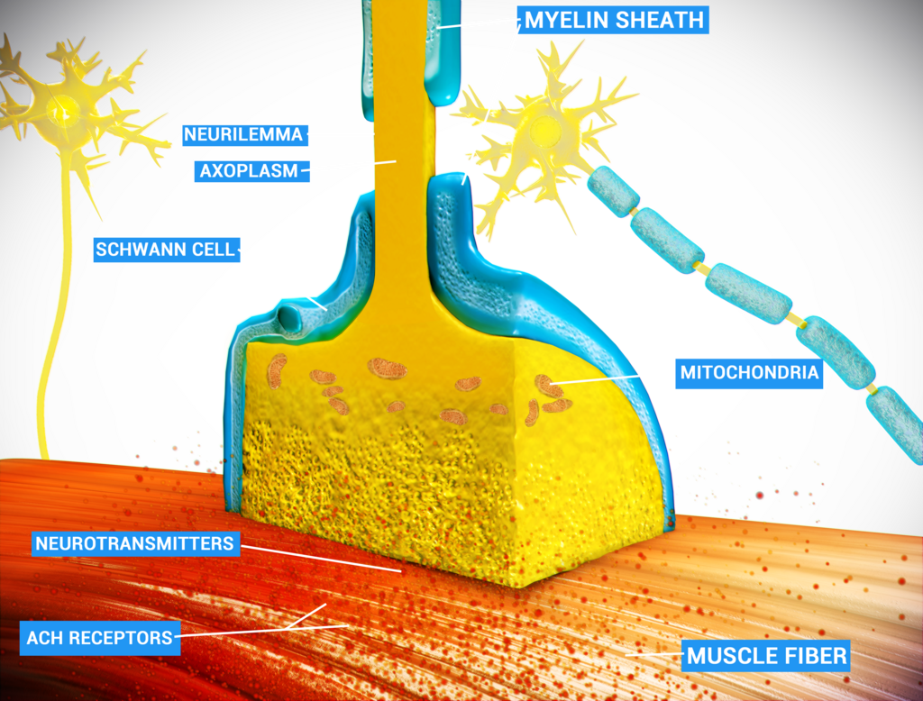
The motor neuron's axon terminal, the presynaptic side, doesn't physically touch the muscle fiber. It hovers just above a specialized region of the muscle membrane called the motor end plate. The tiny gap between them is the synaptic cleft.
When an action potential, the nerve's electrical signal, races down the axon and arrives at the terminal, it triggers the opening of voltage-gated calcium channels. Calcium ions () flood into the terminal. This influx is the crucial trigger that causes synaptic vesicles, tiny sacs filled with the neurotransmitter (ACh), to fuse with the presynaptic membrane and release their contents into the synaptic cleft.
The NMJ is where electric signals, called action potentials, generated by motor neurons in the nerve interact with the muscle, causing the muscle to contract.
The released ACh diffuses across the synaptic cleft in microseconds and binds to nicotinic acetylcholine receptors embedded in the motor end plate. These receptors are ligand-gated ion channels. When ACh binds, the channels open, allowing sodium ions () to rush into the muscle fiber and a smaller amount of potassium ions () to leave. The large influx of positive sodium ions depolarizes the motor end plate, creating a graded potential known as the (EPP). If this EPP reaches the threshold, it triggers a full-blown action potential that spreads across the entire muscle fiber membrane, the sarcolemma.
Inside the Fiber
For a muscle fiber to contract uniformly, the action potential can't just skim along the surface. It needs to penetrate deep inside. The sarcolemma has a solution: a network of invaginations called transverse tubules, or These tunnels carry the action potential from the surface into the fiber's core, ensuring the signal reaches all parts of the cell nearly simultaneously.
The T-tubules run alongside the sarcoplasmic reticulum (SR), a specialized endoplasmic reticulum that serves as an intracellular reservoir for calcium ions. As the action potential travels down a T-tubule, it activates voltage-sensitive proteins embedded in the tubule's membrane called dihydropyridine (DHP) receptors.
In skeletal muscle, the DHP receptors are physically linked to another set of channels on the SR membrane called ryanodine receptors (RyRs). When the action potential causes the DHP receptor to change shape, it mechanically pulls open the ryanodine receptor gate. This is the pivotal moment of excitation-contraction coupling.
The opening of the RyR channels allows the vast stores of calcium ions sequestered inside the SR to flood out into the cytoplasm of the muscle fiber. This sudden, massive increase in intracellular calcium concentration is the direct signal that initiates the contraction of the myofibrils, the protein filaments that do the actual work of the muscle.
What is the primary neurotransmitter responsible for initiating muscle contraction at the neuromuscular junction?
What is the immediate trigger for the release of acetylcholine-filled vesicles from the presynaptic terminal of a motor neuron?
This elegant sequence of events, from an electrical signal in a nerve to a chemical signal at the synapse, back to an electrical signal in the muscle, and finally to the release of calcium, is the fundamental process that allows your brain to command your body to move.