Advanced Male Reproductive Physiology
Seminiferous Tubule Microanatomy
The Sperm Production Factory
The testes are packed with hundreds of tiny, coiled tubes called seminiferous tubules. If you stretched them out, they would span several football pitches. This is where spermatogenesis, the production of sperm, takes place. Think of these tubules as microscopic assembly lines, each with a highly specialised structure to support this complex process.
The main structural cells of these tubules are the Sertoli cells, large cells that extend from the outer edge of the tubule right to the central opening, or lumen. They are the governors of this environment, physically supporting, nourishing, and regulating the developing germ cells, which mature into sperm. These germ cells are nestled between them in various stages of development.
A Protected Space
One of the most critical functions of Sertoli cells is creating the blood-testis barrier. They form with their neighbouring Sertoli cells, creating a seal that divides the tubule into two compartments. This barrier is essential because as sperm cells develop, they produce new proteins on their surface that the body’s immune system would recognise as foreign. Without the barrier, the immune system would attack and destroy the developing sperm.
Tight junctions between these sustentacular cells create the blood–testis barrier, which keeps bloodborne substances from reaching the germ cells and, at the same time, keeps surface antigens on developing germ cells from escaping into the bloodstream and prompting an autoimmune response.
This barrier creates two distinct areas. The basal compartment is on the outer side of the barrier, near the basement membrane. This is where the earliest germ cells, the spermatogonia, reside. As they divide and mature, they must pass through the tight junctions to enter the adluminal compartment, the inner area closer to the lumen. This journey is carefully controlled by the Sertoli cells, ensuring the developing sperm are protected throughout.
The Support Crew
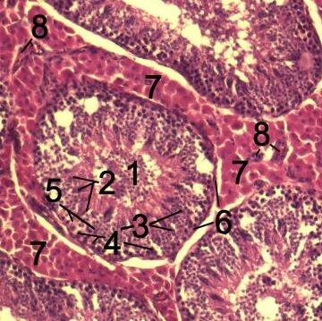
The area outside the seminiferous tubules, known as the interstitial space, is also crucial. It contains blood vessels, connective tissue, and two important cell types.
First are the , which are responsible for producing testosterone. This hormone is vital for spermatogenesis, acting on the Sertoli cells to support germ cell development.
Surrounding each tubule is a thin layer of smooth muscle-like cells called peritubular myoid cells. These cells contract rhythmically, creating gentle waves that help move sperm and fluid through the tubules and out of the testis.
Let's check your understanding of these key anatomical features.
What is the primary function of the seminiferous tubules?
The blood-testis barrier is crucial for protecting developing sperm from the body's immune system. What forms this barrier?
This intricate system of compartments, barriers, and specialised cells ensures a constant and protected supply of new sperm can be produced.